Research at King’s College London has revealed subtle brain differences in adult males with autism spectrum disorder (ASD), which may go some way towards explaining why symptoms persist into adulthood in some people with the disorder.
ASD affects around 1 in 100 people in the UK and involves a spectrum of conditions which manifest themselves differently in different people. People with ASD can have varying levels of impairment across three common areas, which might include: deficits in social interactions and reciprocal understanding, repetitive behaviour and narrow interests, and impairment in language and communication.
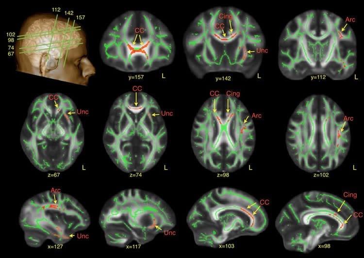
The study, published today in the journal Brain, used a novel brain imaging method to identify altered brain connections in people with ASD. The researchers used Diffusion Tensor Imaging (DTI), a Magnetic Resonance Imaging (MRI) technique, to compare networks of white matter in 61 adults with ASD and 61 controls. White matter consists of large bundles of nerve cells that connect different regions of the brain and enable communication between them.
The scans revealed that men with ASD had differences in brain connections in the frontal lobe, a part of the brain that is crucial to developing language and social interaction skills.
Specifically, these men had altered development of white matter connections in the left side of the brain, the arcuate bundle, which is involved in language. The differences in the arcuate bundle, which connects areas of the brain involved in understanding words and regions related to speech production, were particularly severe in those who had a significant history of ‘delayed echolalia’. Delayed echolalia is very common in ASD and manifests in the parrot-like repetition of words or sentences.
ASD was also associated with underdevelopment of white matter in the left uncinate bundle, which plays a significant role in face recognition and emotional processing. This also correlated with observations of ‘inappropriate use of facial expression’in childhood.

Dr Marco Catani from the Institute of Psychiatry, Psychology & Neuroscience (IoPPN) at King’s College London, said: ‘White matter provides key insights which allow us to paint a precise picture of how different parts of the brain develop during critical periods in childhood.
‘We found subtle brain differences in men who at a very young age had severe problems with communication and social interaction. The differences appear to remain even if they have somehow learned to cope with these difficulties in adult life.
‘It is worth noting that the brain differences are visible only with the special research techniques we now have at our disposal. These differences are very subtle and potentially reversible. Thanks to neuroimaging studies like this, it may one day be possible to stimulate the development of these faulty brain connections, or to predict how people with autism respond to treatment.’
Dr Catani added: ‘Our study did not include women and children, so it would be interesting to explore whether similar differences exist within these groups. For example, research has shown that women appear more resilient than men when it comes to autism, so it will be important if this is explained biologically in their brain development.’
Funding: This research was funded by the Medical Research Council (MRC) as part of the MRC Autism Imaging Multicentre Study (MRC AIMS) Consortium.
Source: Jack Stonebridge – King’s College London
Image Source: The image is credited to the researchers/King’s College London
Original Research: Full open access research for “Frontal networks in adults with autism spectrum disorder” by Marco Catani, Flavio Dell’Acqua, Sanja Budisavljevic, Henrietta Howells, Michel Thiebaut de Schotten, Seán Froudist-Walsh, Lucio D’Anna, Abigail Thompson, Stefano Sandrone, Edward T. Bullmore, John Suckling, Simon Baron-Cohen, Michael V. Lombardo, Sally J. Wheelwright, Bhismadev Chakrabarti, Meng-Chuan Lai, Amber N. V. Ruigrok, Alexander Leemans, Christine Ecker, MRC AIMS Consortium, Michael C. Craig, and Declan G. M. Murphy in Brain. Published online January 29 2016 doi:10.1093/brain/awv351
Abstract
Frontal networks in adults with autism spectrum disorder
It has been postulated that autism spectrum disorder is underpinned by an ‘atypical connectivity’ involving higher-order association brain regions. To test this hypothesis in a large cohort of adults with autism spectrum disorder we compared the white matter networks of 61 adult males with autism spectrum disorder and 61 neurotypical controls, using two complementary approaches to diffusion tensor magnetic resonance imaging. First, we applied tract-based spatial statistics, a ‘whole brain’ non-hypothesis driven method, to identify differences in white matter networks in adults with autism spectrum disorder. Following this we used a tract-specific analysis, based on tractography, to carry out a more detailed analysis of individual tracts identified by tract-based spatial statistics. Finally, within the autism spectrum disorder group, we studied the relationship between diffusion measures and autistic symptom severity. Tract-based spatial statistics revealed that autism spectrum disorder was associated with significantly reduced fractional anisotropy in regions that included frontal lobe pathways. Tractography analysis of these specific pathways showed increased mean and perpendicular diffusivity, and reduced number of streamlines in the anterior and long segments of the arcuate fasciculus, cingulum and uncinate—predominantly in the left hemisphere. Abnormalities were also evident in the anterior portions of the corpus callosum connecting left and right frontal lobes. The degree of microstructural alteration of the arcuate and uncinate fasciculi was associated with severity of symptoms in language and social reciprocity in childhood. Our results indicated that autism spectrum disorder is a developmental condition associated with abnormal connectivity of the frontal lobes. Furthermore our findings showed that male adults with autism spectrum disorder have regional differences in brain anatomy, which correlate with specific aspects of autistic symptoms. Overall these results suggest that autism spectrum disorder is a condition linked to aberrant developmental trajectories of the frontal networks that persist in adult life.
“Frontal networks in adults with autism spectrum disorder” by Marco Catani, Flavio Dell’Acqua, Sanja Budisavljevic, Henrietta Howells, Michel Thiebaut de Schotten, Seán Froudist-Walsh, Lucio D’Anna, Abigail Thompson, Stefano Sandrone, Edward T. Bullmore, John Suckling, Simon Baron-Cohen, Michael V. Lombardo, Sally J. Wheelwright, Bhismadev Chakrabarti, Meng-Chuan Lai, Amber N. V. Ruigrok, Alexander Leemans, Christine Ecker, MRC AIMS Consortium, Michael C. Craig, and Declan G. M. Murphy in Brain. Published online January 29 2016 doi:10.1093/brain/awv351


