Summary: Researchers have, for the first time, tracked how individual neurons lose and recover energy during spreading depolarizations — waves of electrical disturbance linked to brain disorders like stroke. Using genetically modified mice and real-time fluorescent microscopy, the team visualized adenosine triphosphate (ATP) levels in neurons under both healthy and stroke-like conditions.
These waves cause temporary ATP depletion, which worsens during energy deprivation, simulating stroke-induced damage. Importantly, neurons often recovered energy stores once glucose and oxygen were restored, indicating that energy collapse may be reversible.
Key Facts:
- Real-Time Imaging: Neuron ATP depletion was directly visualized during spreading depolarizations.
- Reversible Deficit: Energy loss reversed when glucose and oxygen were reintroduced.
- Stroke-Like Simulation: ATP depletion accelerated dramatically under restricted energy conditions.
Source: University of Leipzig
A research team at the Carl Ludwig Institute for Physiology at Leipzig University has, for the first time, demonstrated how the energy levels of individual neurons in the brain change during so-called spreading depolarizations – waves of activity that occur in various brain disorders.
The findings provide important foundations for understanding energy metabolism in cases of acute cerebral ischaemia, such as that which occurs during a stroke.

The study has just been published in the renowned journal PNAS.
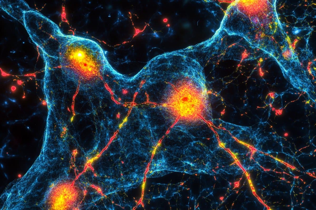
Adenosine triphosphate, or ATP, is an essential energy source in neurons. In the new study, researchers at the Carl Ludwig Institute for Physiology used a specially developed mouse model whose brain neurons produce a fluorescent sensor protein. This allowed them to visualise the available amount of energy in individual neurons.
Using high-resolution fluorescence microscopy, the team was able to observe, in real time, how ATP levels in single neurons changed during spreading depolarizations.
These waves – in which neurons depolarize one after another, much like a short circuit – are linked to progressive tissue damage after stroke. Until now, it was unclear how ATP, the brain’s central energy carrier, behaves in individual neurons during such waves.
“Our study is the first to provide high-resolution insights into how and when neurons lose their energy reserves during an acute mismatch between energy supply and demand, such as in a stroke,” says Dr Karl Schoknecht of the Carl Ludwig Institute for Physiology, lead author of the study.
“Interestingly, the energy reserves are not depleted evenly, but associated with spreading depolarizations. The model will be used in further projects to test potential therapies aimed at preventing the severe energy loss triggered by these waves,” explains the researcher from the Faculty of Medicine.
The findings of the study show that even in ‘healthy’ brain tissue, these waves cause a temporary drop in ATP levels. The effect of spreading depolarizations became particularly pronounced under conditions of energy deprivation – like those that occur during a stroke.
In such cases, the waves greatly accelerated the loss of ATP, leading to the exhaustion of the neurons’ energy reserves. However, even after spreading depolarizations, most neurons were still capable of replenishing their ATP stores – provided that glucose and oxygen were resupplied. This means that the collapse of energy metabolism is, in principle, still reversible.
In this study, the team simulated stroke conditions by removing glucose and oxygen from the nutrient solution. At the same time, they recorded spreading depolarizations using electrophysiological methods. The findings contribute to the understanding of brain energy metabolism.
The study brings together complementary expertise at the Carl Ludwig Institute for Physiology: advanced microscopy from Professor Jens Eilers, the development of specialised mouse models by Professor Johannes Hirrlinger, and Dr Karl Schoknecht’s research on spreading depolarizations.
About this neuroscience research news
Author: Carsten Heckmann
Source: University of Leipzig
Contact: Carsten Heckmann – University of Leipzig
Image: The image is credited to Neuroscience News
Original Research: Closed access.
“Spreading depolarizations exhaust neuronal ATP in a model of cerebral ischemia” by Karl Schoknecht et al. PNAS
Abstract
Spreading depolarizations exhaust neuronal ATP in a model of cerebral ischemia
Spreading depolarizations (SDs) have been identified in various brain pathologies. SDs increase the cerebral energy demand and, concomitantly, oxygen consumption, which indicates enhanced synthesis of adenosine triphosphate (ATP) by oxidative phosphorylation.
Therefore, SDs are considered particularly detrimental during reduced supply of oxygen and glucose. However, measurements of intracellular neuronal ATP ([ATP]i), ultimately reporting the balance of ATP synthesis and consumption during SDs, have not yet been conducted.
Here, we investigated neuronal ATP homeostasis during SDs using two-photon imaging in acute brain slices from adult mice expressing the ATP sensor ATeam1.03YEMK in neurons. SDs were induced by application of potassium chloride or by oxygen and glucose deprivation (OGD) and detected by recording the local field potential, extracellular potassium, as well as the intrinsic optical signal.
We found that, in the presence of oxygen and glucose, SDs were accompanied by a substantial but transient drop in neuronal ATP sensor signals, corresponding to a drop in ATP. OGD, which prior to SDs was accompanied by only a slight reduction in ATP signals, led to a large, terminal drop in ATP signals during SDs.
Subsequently, we investigated whether neurons could still regenerate ATP if oxygen and glucose were promptly resupplied following SD detection, and show that ATP depletion was essentially reversible in most cells. Our findings indicate that SDs are accompanied by a substantial increase in ATP consumption beyond production.
This, under conditions that mimic reduced blood supply, leads to a breakdown of [ATP]i. Therefore, our findings support therapeutic strategies targeting SDs after cerebral ischemia.


