

Summary: Researchers have identified a group of neurons that relay information from the brain’s respiratory system to an areas of the brain associated with generating arousal.
Source: Stanford.
Stanford scientists have identified a small group of neurons that communicates goings-on in the brain’s respiratory control center to the structure responsible for generating arousal throughout the brain.
Try it. Breathe slowly and smoothly. A pervasive sense of calm descends. Now breathe rapidly and frenetically. Tension mounts. Why?
It’s a question that has never been answered by science, until now.
In a new study, researchers at the Stanford University School of Medicine and their colleagues have identified a handful of nerve cells in the brainstem that connect breathing to states of mind.
A paper describing the findings were published March 31 in Science. Mark Krasnow, MD, PhD, professor of biochemistry, is the senior author. The lead author is former Stanford graduate student Kevin Yackle, MD, PhD, now a faculty fellow at the University of California-San Francisco.
Medical practitioners sometimes prescribe breathing-control exercises for people with stress disorders. Similarly, the practice of pranayama — controlling breath in order to shift one’s consciousness from an aroused or even frantic state to a more meditative one — is a core component of virtually all varieties of yoga.
“This study is intriguing because it provides a cellular and molecular understanding of how that might work,” Krasnow said.
Tiny cluster of neurons
The tiny cluster of neurons linking respiration to relaxation, attention, excitement and anxiety is located deep in the brainstem. This cluster, located in an area Krasnow calls the pacemaker for breathing, was discovered in mice by study co-author Jack Feldman, PhD, a professor of neurobiology at UCLA, who published his findings in 1991. An equivalent structure has since been identified in humans.
“The respiratory pacemaker has, in some respects, a tougher job than its counterpart in the heart,” said Krasnow, who is also a Howard Hughes Medical Institute investigator. “Unlike the heart’s one-dimensional, slow-to-fast continuum, there are many distinct types of breaths: regular, excited, sighing, yawning, gasping, sleeping, laughing, sobbing. We wondered if different subtypes of neurons within the respiratory control center might be in charge of generating these different types of breath.”
On that hunch, Yackle searched through public databases to assemble a list of genes that are preferentially activated in the part of the mouse brainstem where the breathing-control center resides. This center’s technical term is the pre-Bötzinger complex, or preBötC.
He pinpointed a number of such genes, allowing the investigators to identify more than 60 separate neuronal subtypes, physically differentiated from one another by their gene-activation signatures but comingling in the preBötC like well-stirred spaghetti strands. The scientists were able to use these genes, and the protein products for which they are recipes, as markers allowing them to zero in on the different neuronal subtypes.
Knocking out neurons
Now the scientists could systematically assess the role of each neuronal subpopulation in laboratory mice. With advanced technologies, they could selectively destroy any one of these neuronal subtypes — and only that subtype — based on its unique signature of active genes. Then they could observe how this particular subtype’s loss affected the animals’ breathing. In 2016, in collaboration with Feldman, they succeeded in isolating a subpopulation of neurons in the preBötC that explicitly controls one type of breathing: sighing. Knocking out these neurons eliminated sighing but left other modes of breathing unaffected. The discovery was published in Nature in 2016.
Krasnow and Yackle then set out to discover the respiratory role of another subpopulation of about 175 preBötC neurons distinguished by their shared expression of two genetic markers called Cdh9 and Dbx1. They bioengineered mice in which they could wipe out, at will, the neurons bearing both of these markers.
But once these rodents had their Cdh9/Dbx1 neurons eliminated, they seemed to take the loss in stride. Unlike their sigh-deprived brethren, there was no lacuna in these mice’s portfolio of breathing variations.
“I was initially disappointed,” said Yackle.
But a few days afterward, he noticed something: For mice, the animals were extraordinarily calm. “If you put them in a novel environment, which normally stimulates lots of sniffing and exploration,” Yackle said, “they would just sit around grooming themselves” — evidence of what passes for mellowness when you’re a mouse.
Chilling out
Further analysis showed that while these mice still displayed the full palette of breathing varieties from sighs to sniffs, the relative proportions of those varieties had changed. There were fewer fast “active” and faster “sniffing” breaths, and more slow breaths associated with chilling out.
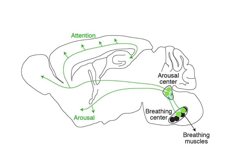
The investigators surmised that rather than regulating breathing, these neurons were spying on it instead and reporting their finding to another structure in the brainstem. This structure, the locus coeruleus, sends projections to practically every part of the brain and drives arousal: waking us from sleep, maintaining our alertness and, if excessive, triggering anxiety and distress. It’s known that neurons in the locus coeruleus exhibit rhythmic behavior whose timing is correlated with that of breathing. In a series of experiments, the Stanford researchers proved that the preBötC neurons that express Cadh9 and Dbx1 not only project to the locus coeruleus — a new finding — but activate its long-distance-projections, promoting brainwide arousal.
“If something’s impairing or accelerating your breathing, you need to know right away,” said Krasnow. “These 175 neurons, which tell the rest of the brain what’s going on, are absolutely critical.”
“The preBötC now appears to play a key role in the effects of breathing on arousal and emotion, such as seen during meditation,” said Feldman. “We’re hopeful that understanding this center’s function will lead to therapies for stress, depression and other negative emotions.”
Other Stanford co-authors are John Huguenard, PhD, professor of neurology and neurological sciences; Liqun Luo, PhD, professor of biology and an HHMI investigator; former postdoctoral scholar Lindsay Schwarz, PhD; and graduate student Jordan Sorkin.
A researcher at the Chicago Medical School also co-authored the study.
Krasnow is also executive director of the Wall Center for Pulmonary Vascular Disease, a member of the Stanford’s Neurosciences Institute, Cardiovascular Institute, Cancer Institute and Bio-X.
Funding: The study was funded by the National Institutes of Health (grants HL70029 and HL40959) and HHMI.
Stanford’s Department of Biochemistry also supported the work.
Source: Bruce Goldman- Stanford
Image Source: NeuroscienceNews.com image is credited to Krasnow lab.
Original Research: Abstract for “Breathing control center neurons that promote arousal in mice” by Kevin Yackle, Lindsay A. Schwarz, Kaiwen Kam, Jordan M. Sorokin, John R. Huguenard, Jack L. Feldman, Liqun Luo, and Mark A. Krasnow in Science. Published online March 31 2017 doi:10.1126/science.aai7984
[cbtabs][cbtab title=”MLA”]Stanford “How Slow Breathing Induces Tranquility.” NeuroscienceNews. NeuroscienceNews, 31 March 2017.
<https://neurosciencenews.com/tranquility-slow-breathing-6317/>.[/cbtab][cbtab title=”APA”]Stanford (2017, March 31). How Slow Breathing Induces Tranquility. NeuroscienceNew. Retrieved March 31, 2017 from https://neurosciencenews.com/tranquility-slow-breathing-6317/[/cbtab][cbtab title=”Chicago”]Stanford “How Slow Breathing Induces Tranquility.” https://neurosciencenews.com/tranquility-slow-breathing-6317/ (accessed March 31, 2017).[/cbtab][/cbtabs]
Abstract
Breathing control center neurons that promote arousal in mice
Slow, controlled breathing has been used for centuries to promote mental calming, and it is used clinically to suppress excessive arousal such as panic attacks. However, the physiological and neural basis of the relationship between breathing and higher-order brain activity is unknown. We found a neuronal subpopulation in the mouse preBötzinger complex (preBötC), the primary breathing rhythm generator, which regulates the balance between calm and arousal behaviors. Conditional, bilateral genetic ablation of the ~175 Cdh9/Dbx1 double-positive preBötC neurons in adult mice left breathing intact but increased calm behaviors and decreased time in aroused states. These neurons project to, synapse on, and positively regulate noradrenergic neurons in the locus coeruleus, a brain center implicated in attention, arousal, and panic that projects throughout the brain.
“Breathing control center neurons that promote arousal in mice” by Kevin Yackle, Lindsay A. Schwarz, Kaiwen Kam, Jordan M. Sorokin, John R. Huguenard, Jack L. Feldman, Liqun Luo, and Mark A. Krasnow in Science. Published online March 31 2017 doi:10.1126/science.aai7984


