

Summary: Researchers believe they may have pinpointed an area of the brain that plays a role in maintaining human consciousness.
Source: Beth Israel Deaconess Medical Center.
Study reveals a network within the brain that plays a role maintaining consciousness.
Philosophers have long struggled to define human consciousness. Now, a team of researchers led by neurologists at Beth Israel Deaconess Medical Center (BIDMC) has pinpointed the regions of the brain that may play a role maintaining it. Their findings, which have already garnered multiple awards from the American Academy of Neurology, were published today in that society’s journal, Neurology.
“For the first time, we have found a connection between the brainstem region involved in arousal and regions involved in awareness, two prerequisites for consciousness,” said Michael D. Fox, MD, PhD, Director of the Laboratory for Brain Network Imaging and Modulation and the Associate Director of the Berenson-Allen Center for Noninvasive Brain Stimulation at BIDMC. “A lot of pieces of evidence all came together to point to this network playing a role in human consciousness.”
Classical neurology holds that arousal and awareness are two critical components of consciousness. Arousal is likely regulated by the brainstem – the portion of the brain, contiguous with the spinal cord, that is responsible for the sleep/wake cycle and cardiac and respiratory rates. Awareness, another critical component of consciousness, has long been thought to reside somewhere in the cortex, the outer layer of the brain responsible for many of its higher functions.
The researchers analyzed 36 patients with brainstem lesions, of which 12 led to coma and 24 did not. Mapping the injuries revealed that a small “coma-specific” area of the brainstem – the rostral dorsolateral pontine tegmentum – was significantly associated with coma. Ten out of the 12 coma-inducing brainstem lesions involved this area, while just one of the 24 control lesions did.
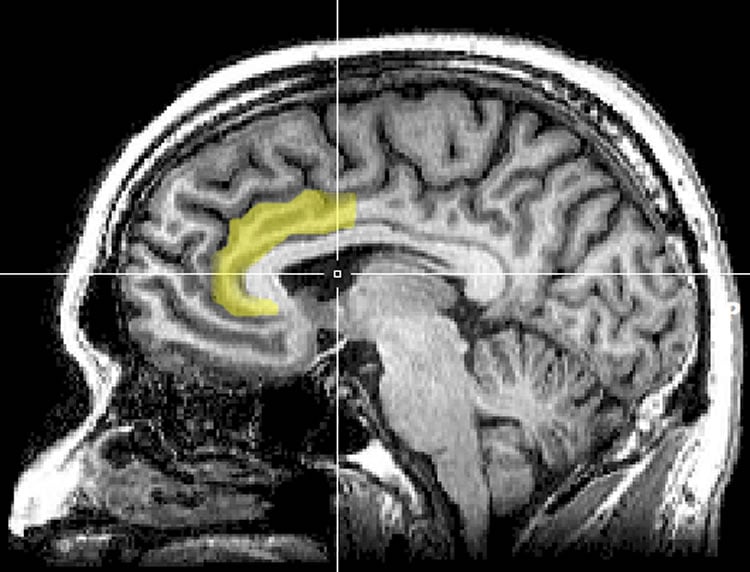
Armed with that information, Fox and colleagues, including lead author David Fischer, MD, then a medical student at Harvard Medical School, used a wiring diagram of the healthy human brain – based on a large, shared data set called the Human Connectome – to identify which other parts of the brain were connected to these coma-causing lesions. Their analysis revealed two areas in the cortex of the brain that were significantly connected to the coma-specific region of the brainstem. One sat in the left, ventral, anterior insula (AI), the other in the pregenual anterior cingulate cortex (pACC). Both regions have been implicated previously in arousal and awareness.
“We now have a great map of how the brain is wired up in the Human Connectome,” said Fox, who is also an Assistant Professor of Neurology at Harvard Medical School. “We can look at not just the location of lesions, but also their connectivity. Over the past year, researchers in my lab have used this approach to understand visual and auditory hallucinations, impaired speech, and movement disorders. A collaborative team of neuroscientists and physicians had the insight and unique expertise needed to apply this approach to consciousness.”
The team included co-lead author, Aaron Boes, MD, PhD, and co-senior author, Joel Geerling, MD, PhD, both formerly of BIDMC and now of University of Iowa Carver College of Medicine.
Finally, the team investigated whether this brainstem-cortex network was functioning in another subset of patients with disorders of consciousness, including coma. Using a special type of MRI scan, the scientists found that their newly identified “consciousness network” was disrupted in patients with impaired consciousness. The findings – bolstered by data from rodent studies – suggest the network between the brainstem and these two cortical regions plays a role maintaining human consciousness.
“The added value of thinking about coma as a network disorder is it presents possible targets for therapy, such as using brain stimulation to augment recovery,” Boes said.
A next step, Fox notes, may be to investigate other data sets in which patients lost consciousness to find out if the same, different or overlapping neural networks are involved.
“This is most relevant if we can use these networks as a target for brain stimulation for people with disorders of consciousness,” said Fox. “If we zero in on the regions and network involved, can we someday wake someone up who is in a persistent vegetative state? That’s the ultimate question.”
Study coauthors include David B. Fischer, MD, Aaron D. Boes, MD, PhD, Joel C. Geerling, MD, PhD, Clifford B. Saper, MD, PhD, and Alvaro Pascual-Leone, MD, PhD, of BIDMC; Athena Demertzi, PhD, of the Brain and Spine Institute (Institut du Cerveau et de la Moelle épinière-ICM), Hôpital Pitié-Salpêtrière, Paris, France and the Coma Science Group, GIGA-Research & Cyclotron Research Centre, University and University Hospital of Liège, Belgium; Steven Laureys, PhD, also of the Coma Science Group; Henry C. Evrard, PhD, of the Functional and Comparative Neuroanatomy Lab and the Centre for Integrative Neuroscience, Tübingen and the Max Planck Institute for Biological Cybernetics, Tübingen, Germany; Brian L. Edlow, MD, and Hesheng Liu, PhD. of the Athinoula A. Martinos Center for Biomedical Imaging at Massachusetts General Hospital, Charlestown, MA.
Funding: This work was supported by the Howard Hughes Medical Institute, the Parkinson’s Disease Foundation, the NIH (Shared Instrument Grant S10RR023043, K23NS083741, R01HD069776, R01NS073601, R01NS085477, R21MH099196, R21NS082870, R21NS085491, R21HD07616, R25NS065743, R25NS070682, T32 HL007901, P01HL095491), American Academy of Neurology/American Brain Foundation, Sidney R. Baer, Jr. Foundation, Harvard Catalyst, the Belgian National Funds for Scientific Research, the European Commission, the James McDonnell Foundation, the European Space Agency, Mind Science Foundation, the French Speaking Community Concerted Research Action (ARC-06/11-340), the Public Utility Foundation “Université Européenne du Travail,” “Fondazione Europea di Ricerca Biomedica,” the University and University Hospital of Liège, the Center for Integrative Neuroscience, and the Max Planck Society.
Source: Beth Israel Deaconess Medical Center
Image Source: This NeuroscienceNews.com image is adapted from the CMU press release.
Original Research: Abstract for “A human brain network derived from coma-causing brainstem lesions” by David B. Fischer, Aaron D. Boes, Athena Demertzi, Henry C. Evrard, Steven Laureys, Brian L. Edlow, Hesheng Liu, Clifford B. Saper, Alvaro Pascual-Leone, Michael D. Fox, and Joel C. Geerling in Neurology. Published online November 4 2016 doi:10.1212/WNL.0000000000003404
[cbtabs][cbtab title=”MLA”]Beth Israel Deaconess Medical Center. “New Theory Debunks Idea That Math Abilities Are Inate.” NeuroscienceNews. NeuroscienceNews, 5 November 2016.
<https://neurosciencenews.com/consciousness-neuroscience-pacc-5436/>.[/cbtab][cbtab title=”APA”]Beth Israel Deaconess Medical Center. (2016, November 5). New Theory Debunks Idea That Math Abilities Are Inate. NeuroscienceNews. Retrieved November 5, 2016 from https://neurosciencenews.com/consciousness-neuroscience-pacc-5436/[/cbtab][cbtab title=”Chicago”]Beth Israel Deaconess Medical Center. “New Theory Debunks Idea That Math Abilities Are Inate.” https://neurosciencenews.com/consciousness-neuroscience-pacc-5436/ (accessed November 5, 2016).[/cbtab][/cbtabs]
Abstract
A human brain network derived from coma-causing brainstem lesions
Objective: To characterize a brainstem location specific to coma-causing lesions, and its functional connectivity network.
Methods: We compared 12 coma-causing brainstem lesions to 24 control brainstem lesions using voxel-based lesion-symptom mapping in a case-control design to identify a site significantly associated with coma. We next used resting-state functional connectivity from a healthy cohort to identify a network of regions functionally connected to this brainstem site. We further investigated the cortical regions of this network by comparing their spatial topography to that of known networks and by evaluating their functional connectivity in patients with disorders of consciousness.
Results: A small region in the rostral dorsolateral pontine tegmentum was significantly associated with coma-causing lesions. In healthy adults, this brainstem site was functionally connected to the ventral anterior insula (AI) and pregenual anterior cingulate cortex (pACC). These cortical areas aligned poorly with previously defined resting-state networks, better matching the distribution of von Economo neurons. Finally, connectivity between the AI and pACC was disrupted in patients with disorders of consciousness, and to a greater degree than other brain networks.
Conclusions: Injury to a small region in the pontine tegmentum is significantly associated with coma. This brainstem site is functionally connected to 2 cortical regions, the AI and pACC, which become disconnected in disorders of consciousness. This network of brain regions may have a role in the maintenance of human consciousness.
“A human brain network derived from coma-causing brainstem lesions” by David B. Fischer, Aaron D. Boes, Athena Demertzi, Henry C. Evrard, Steven Laureys, Brian L. Edlow, Hesheng Liu, Clifford B. Saper, Alvaro Pascual-Leone, Michael D. Fox, and Joel C. Geerling in Neurology. Published online November 4 2016 doi:10.1212/WNL.0000000000003404


