

Understanding how the Christmas spirit works could be a powerful tool in treating the ‘bah humbug’ syndrome.
The Christmas spirit has been located in the human brain, reveals a study published in The BMJ‘s Christmas issue this week.
The Christmas spirit has been a widespread phenomenon for centuries and is commonly described as feelings of joy and nostalgia mixed with associations to merry feelings, gifts, delightful smells, and good food.
However, the authors of the study estimate that “millions of people are prone to displaying Christmas spirit deficiencies,” and refer to this as the ‘bah humbug’ syndrome.
“Accurate localisation of the Christmas spirit is a paramount first step in being able to help this group of patients,” they say, and can advance “understanding of the brain’s role in festive cultural traditions.”
So the team of researchers from Rigshospitalet, a hospital affiliated with Copenhagen University, attempted to locate the ‘Christmas spirit’ in the brain using functional magnetic resonance imaging (fMRI).
An fMRI scan measures changes in blood oxygenation and flow that occur in response to neural activity, and can produce activation maps showing which parts of the brain are involved in a particular mental process.
The study involved 10 participants who celebrated Christmas, and 10 healthy participants who lived in the same area, but who had no Christmas traditions.
All participants were healthy, and did not consume eggnog or gingerbread before the scans.
Each participant was scanned while they viewed 84 images with video goggles.
Images were displayed for two seconds each, and after six consecutive images with a Christmas theme, there were six every day images (see link below for examples).
After the scans, all participants filled out a questionnaire about their Christmas traditions, feelings associated with Christmas, and ethnicity.
Based on these results, 10 participants were allocated to the ‘Christmas group’ — those who celebrated Christmas and had positive associations — and 10 to the ‘non-Christmas group’ — those who did not celebrate Christmas and had neutral feelings towards the festivities.
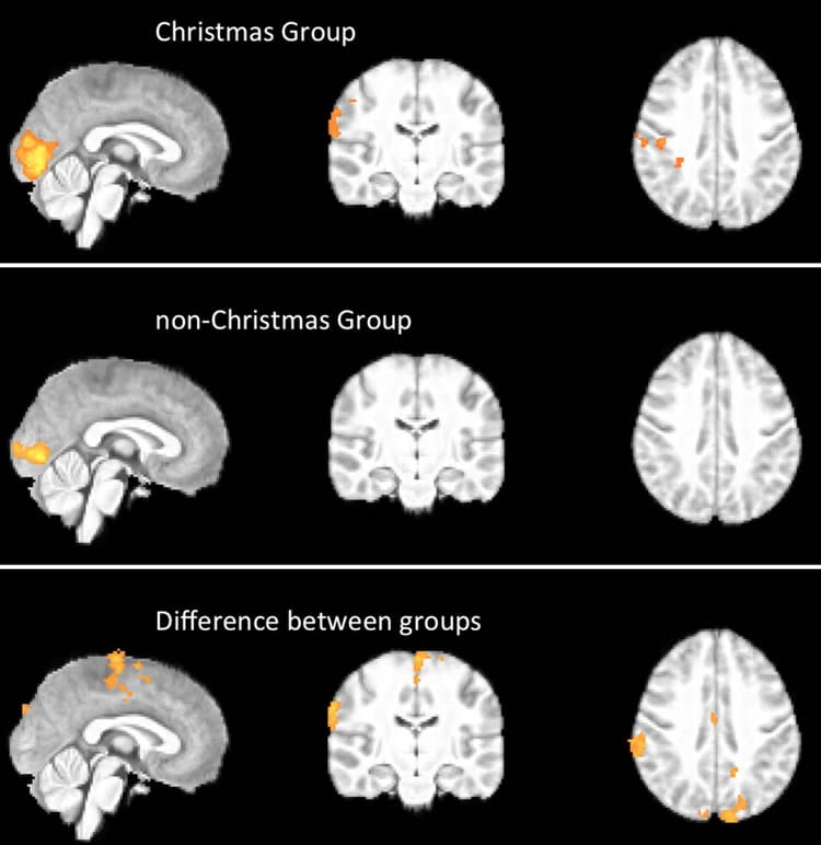
Differences in the brain activation maps from the scans of the two groups were analysed to identify Christmas specific brain activation.
Results showed five areas where the Christmas group responded to Christmas images with a higher activation than the non-Christmas group.
These included the left primary motor and premotor cortex, right inferior and superior parietal lobule, and bilateral primary somatosensory cortex.
These cerebral areas have been associated with spirituality, somatic senses, and recognition of facial emotion among many other functions.
For example, the left and right parietal lobules have been shown to play a role in self transcendence, the personality trait regarding predisposition to spirituality.
In addition, the frontal premotor cortex is important for experiencing emotions shared with others by mirroring or copying their body state, and premotor cortical mirror neurons even respond to observation of ingestive mouth actions.
Further research is necessary to understand the Christmas spirit, and other potential holiday circuits in the brain, say the authors, such as Easter, Chanukah, Eid, and Diwali.
“Although merry and intriguing, these findings should be interpreted with caution,” they explain. “Something as magical and complex as the Christmas spirit cannot be fully explained by, or limited to, the mapped brain activity alone.”
Source: Gozde Zorlu – BMJ
Image Credit: The image is credited to Hougaard et al./BMJ
Original Research: Full open access research for “Evidence of a Christmas spirit network in the brain: functional MRI study” by Anders Hougaard, Ulrich Lindberg, Nanna Arngrim, Henrik B W Larsson, Jes Olesen, Faisal Mohammad Amin, Messoud Ashina, and Bryan T Haddock in BMJ. Published online December 16 2015 doi:10.1136/bmj.h6266
Abstract
Evidence of a Christmas spirit network in the brain: functional MRI study
Objective To detect and localise the Christmas spirit in the human brain.
Design Single blinded, cross cultural group study with functional magnetic resonance imaging (fMRI).
Setting Functional imaging unit and department of clinical physiology, nuclear medicine and PET in Denmark.
Participants 10 healthy people from the Copenhagen area who routinely celebrate Christmas and 10 healthy people living in the same area who have no Christmas traditions.
Main outcome measures Brain activation unique to the group with Christmas traditions during visual stimulation with images with a Christmas theme.
Methods Functional brain scans optimised for detection of the blood oxygen level dependent (BOLD) response were performed while participants viewed a series of images with Christmas themes interleaved with neutral images having similar characteristics but containing nothing that symbolises Christmas. After scanning, participants answered a questionnaire about their Christmas traditions and the associations they have with Christmas. Brain activation maps from scanning were analysed for Christmas related activation in the “Christmas” and “non-Christmas” groups individually. Subsequently, differences between the two groups were calculated to determine Christmas specific brain activation.
Results Significant clusters of increased BOLD activation in the sensory motor cortex, the premotor and primary motor cortex, and the parietal lobule (inferior and superior) were found in scans of people who celebrate Christmas with positive associations compared with scans in a group having no Christmas traditions and neutral associations. These cerebral areas have been associated with spirituality, somatic senses, and recognition of facial emotion among many other functions.
Conclusions There is a “Christmas spirit network” in the human brain comprising several cortical areas. This network had a significantly higher activation in a people who celebrate Christmas with positive associations as opposed to a people who have no Christmas traditions and neutral associations. Further research is necessary to understand this and other potential holiday circuits in the brain. Although merry and intriguing, these findings should be interpreted with caution.
“Evidence of a Christmas spirit network in the brain: functional MRI study” by Anders Hougaard, Ulrich Lindberg, Nanna Arngrim, Henrik B W Larsson, Jes Olesen, Faisal Mohammad Amin, Messoud Ashina, and Bryan T Haddock in BMJ. Published online December 16 2015 doi:10.1136/bmj.h6266


