Summary: A novel X-ray scintillator-based optogenetics technique allows researchers to control neural function deep within the brain and alter behavioral responses.
Source: Fujita Health University School of Medicine
Conventional optogenetics involves invasive implantation of optical fibers in target brain tissues. This is especially challenging for deep areas of the brain owing to extensive tissue damage.
A group of neuroscientists and material scientists now reveal a novel X-ray and scintillator based-optogenetics technique that allows control of neural function deep in the brain without causing any damage. This study adds another aspect to the long list of uses of X-rays in biology and medicine.
Optogenetics is an elegant technique of controlling neurons in the brain using light. Neurons are loaded with special light-sensitive proteins called opsins that sense light (in the visible range), convert it into electric signals, and activate neurons.
Conventionally, optical fibers connected to a light source are invasively implanted into the target tissue. But reaching the deep areas of the brain using this technique is usually accompanied by extensive tissue damage, light toxicity, and harmful effects of thermal irradiation.
To reduce this damage, scientists use microparticles that emit visible light in response to near-infrared irradiation (NIR). These particles are injected into the target tissue and neuron activation is achieved without tissue damage. However, NIR has its limitations—it can only penetrate some millimeters of tissue.
Now, researchers from Japan have finally overcome this challenge by using X-rays to penetrate deep regions of the brain. The findings of this multidisciplinary study, all set to be published in Nature Communications, was led by neuroscientists Prof. Takayuki Yamashita and Dr. Takanori Matsubara from the Department of Physiology, Fujita Health University School of Medicine, and material scientist Dr. Takayuki Yanagida from Nara Institute of Science and Technology.
Prof. Yamashita explains the use of X-rays in their study: “This new technology enables remote control of brain functions in living animals without damaging radio-sensitive cells in the body.”
X-rays are widely used for imaging the human body because of their penetrative capacity. Using this ability of the X-rays, the researchers could reach the deeper areas of the brain. However, opsins do not respond to X-rays. So, to convert the radiation into visible light, the researchers chose materials called scintillators.
Prof. Yamashita explains, “Scintillators, which emit visible light when irradiated with X-rays, have widely been used in X-ray inspection machines and CT scans, but their applications in biology have been limited. This work is the first application of scintillators to behavioral neuroscience.”
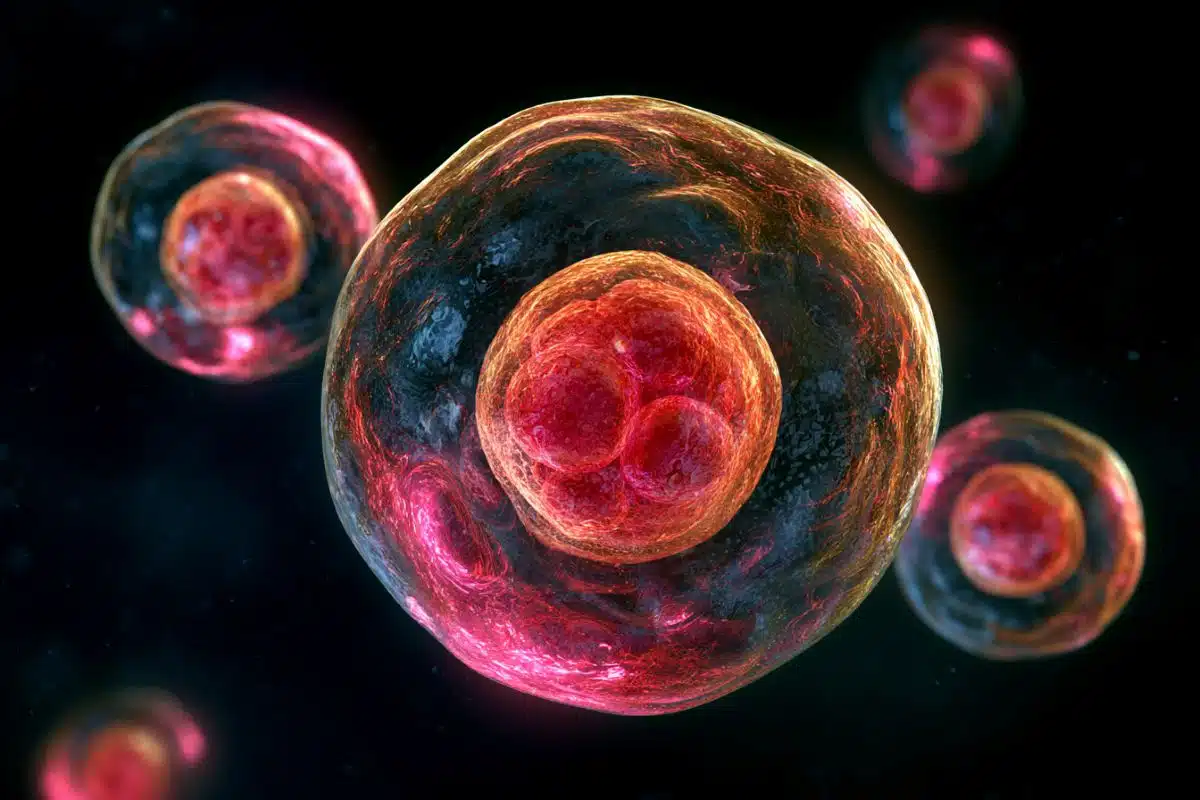
The researchers crushed yellow-emitting inorganic scintillator crystals synthesized in the lab and injected the microparticles into mice brain cells. When X-rays were irradiated onto a dissected mouse head, they passed through skin, skull, and brain tissue. In response to the X-rays, the microparticles emitted yellow light which activated the opsins for excitation and inhibition of neurons. The researchers were able to confirm these results in live mice.

To explore further, the researchers also tested whether the control of neural function by X-rays and opsins could induce a change in behavior. For this, they performed a conditioned place preference test in which mice were given a choice between two compartments, one out of which was exposed to X-ray radiation.
The mice either had excitatory opsins or inhibitory opsins in neurons that govern this type of behavior. Before X-ray irradiation, no mice showed any preference for a compartment. However, after X-ray irradiation, mice with excitatory opsins had an increased preference for the X-ray irradiated compartment and mice with inhibitory opsins showed the opposite behavior.
These results show the advantages of X-rays over conventional methods of optogenetics.
Prof. Yamashita is excited by the achievement of the team, “There are many biomedical technologies that use light to control protein functions. However, these technologies have not been easy to apply to deep tissues owing to the low penetration of stimulating light into the tissue. In contrast, our technology is almost unconstrained by the depth of the tissue.” It also helps that scintillators are bio-compatible, do not cause any tissue damage, and are non-toxic even for long-term implantation.
Scintillator-based optogenetics has many potential applications, from modulating neurons for research to treatment of neurological disorders. The non-invasive nature of this technique allows experiments to be conducted without wires and plugs, not hindering observation and science.
The world of neuroexploration thus seems to have found a new and wonderful probing technique.
About this neurotech research news
Neuroscience News would like to thank Brijesh Manek for submitting this research news for inclusion.
Source: Fujita Health University School of Medicine
Contact: Brijesh Manek – Fujita Health University School of Medicine
Image: The image is credited to Takayuki Yamashita from Fujita Health University
Original Research: Open access.
“Remote control of neural function by X-ray-induced scintillation” by Takanori Matsubara, Takayuki Yanagida, Noriaki Kawaguchi, Takashi Nakano, Junichiro Yoshimoto, Maiko Sezaki, Hitoshi Takizawa, Satoshi P. Tsunoda, Shin-ichiro Horigane, Shuhei Ueda, Sayaka Takemoto-Kimura, Hideki Kandori, Akihiro Yamanaka & Takayuki Yamashita. Nature Communications
Abstract
Remote control of neural function by X-ray-induced scintillation
Scintillators emit visible luminescence when irradiated with X-rays. Given the unlimited tissue penetration of X-rays, the employment of scintillators could enable remote optogenetic control of neural functions at any depth of the brain.
Here we show that a yellow-emitting inorganic scintillator, Ce-doped Gd3(Al,Ga)5O12 (Ce:GAGG), can effectively activate red-shifted excitatory and inhibitory opsins, ChRmine and GtACR1, respectively. Using injectable Ce:GAGG microparticles, we successfully activated and inhibited midbrain dopamine neurons in freely moving mice by X-ray irradiation, producing bidirectional modulation of place preference behavior. Ce:GAGG microparticles are non-cytotoxic and biocompatible, allowing for chronic implantation.
Pulsed X-ray irradiation at a clinical dose level is sufficient to elicit behavioral changes without reducing the number of radiosensitive cells in the brain and bone marrow.
Thus, scintillator-mediated optogenetics enables minimally invasive, wireless control of cellular functions at any tissue depth in living animals, expanding X-ray applications to functional studies of biology and medicine.