

Our brains are constantly barraged with sensory information, but have an amazing ability to filter out just what they need to understand what’s going on around us. For instance, if you stand perfectly still in a room, and that room rotates around you, it’s terrifying. But stand still in a room and turn your eyes, and the same visual input feels perfectly normal. That’s thanks to a complex process in our brain that tell us when and how to pay attention to sensory input. Specifically, we ignore visual input caused by our own eye movements.
Now, researchers at The Rockefeller University have identified a similar process in flies, whose brains ignore visual input caused by their own flight turns. This advance will allow researchers to better understand how ongoing behavior influences visual perception.
“Fly brains are small, so discovering that flies can ‘silence’ visual inputs means that we can aim for a comprehensive understanding of how this silencing process is implemented,” says study author Gaby Maimon, head of the Laboratory of Integrative Brain Function at Rockefeller. Postdoctoral fellows Anmo J. Kim and Jamie K. Fitzgerald, alongside Maimon, report their findings in the August issue of Nature Neuroscience.
By studying the details of this silencing mechanism in flies, we will gain a better understanding of how what we do influences what we see, Maimon says. These mechanisms are disrupted in people with schizophrenia, who have trouble interpreting sensory input properly.
In 1950, researchers Erich von Holst and Horst Mittelstaedt performed a simple experiment: They rotated the environment surrounding flies, and saw they walked in circles in response. But when flies perform a normal turn while walking, they also experience a similarly rotating stimulus on their retina, which they happily ignore. The researchers postulated that the brain must contain a mechanism that tells organisms to ignore what they see when they rotate their eyes or bodies. But the scientists didn’t have the technology to prove it.
To determine if silencing does, in fact, happen in flies, Kim, Fitzgerald, and Maimon attached flies to a tiny harness that allows the animals to flap their wings in tethered flight– the flying equivalent of walking on a treadmill — while the experimenters measured electrical activity from neurons. When these flies attempted to turn left or right, the researchers saw electrical signals that indicated the motor part of the brain was briefly silencing the visual part, says Maimon says. “In simple terms, the motor part of the brain is telling the visual part ‘don’t pay attention to this information, this is not relevant to anything that’s happening in the world.'”
Scientists have seen signatures of the same process in primate brains, but because humans and other primates have billions of neurons, it’s difficult to understand the precise mechanisms involved, Maimon says. “Since fly brains contain 100,000 times fewer neurons than human brains, we should be able to develop a much deeper understanding of this process, and build a blueprint for how to think about sensory silencing across all animals, and, ultimately, even humans.”
Source: Wynne Parry – Rockefeller University
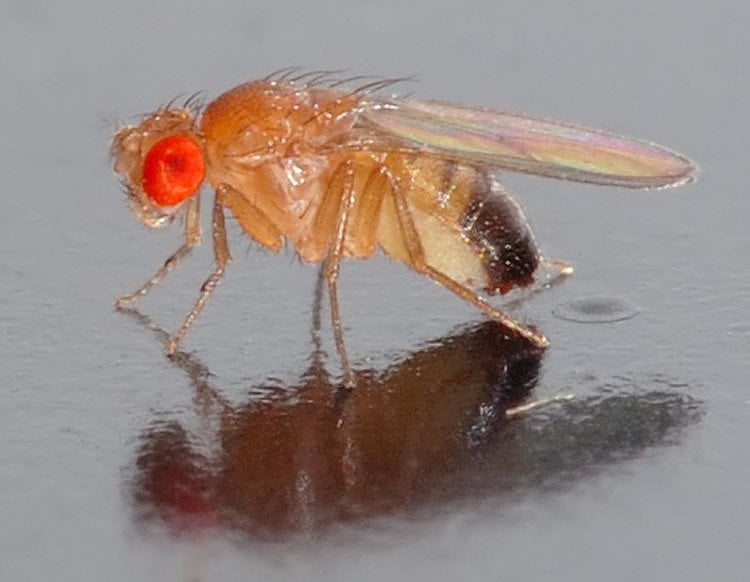
Image Credit: The image is credited to André Karwath and is licensed CC BY SA 2.5
Original Research: Abstract for “Cellular evidence for efference copy in Drosophila visuomotor processing” by Anmo J Kim, Jamie K Fitzgerald and Gaby Maimon in Nature Neuroscience. Published online August 3 2015 doi:10.1038/nn.4083
Abstract
Cellular evidence for efference copy in Drosophila visuomotor processing
Each time a locomoting fly turns, the visual image sweeps over the retina and generates a motion stimulus. Classic behavioral experiments suggested that flies use active neural-circuit mechanisms to suppress the perception of self-generated visual motion during intended turns. Direct electrophysiological evidence, however, has been lacking. We found that visual neurons in Drosophila receive motor-related inputs during rapid flight turns. These inputs arrived with a sign and latency appropriate for suppressing each targeted cell’s visual response to the turn. Precise measurements of behavioral and neuronal response latencies supported the idea that motor-related inputs to optic flow–processing cells represent internal predictions of the expected visual drive induced by voluntary turns. Motor-related inputs to small object–selective visual neurons could reflect either proprioceptive feedback from the turn or internally generated signals. Our results in Drosophila echo the suppression of visual perception during rapid eye movements in primates, demonstrating common functional principles of sensorimotor processing across phyla.
“Cellular evidence for efference copy in Drosophila visuomotor processing” by Anmo J Kim, Jamie K Fitzgerald and Gaby Maimon in Nature Neuroscience. Published online August 3 2015 doi:10.1038/nn.4083


