Summary: A new study reports prolonged exposure to stress can result in structural changes to the amygdala in mice.
Source: Rockefeller University.
Chronic stress can make us worn-out, anxious, depressed–in fact, it can change the architecture of the brain. New research at The Rockefeller University shows that when mice experience prolonged stress, structural changes occur within a little-studied region of their amygdala, a part of the brain that regulates basic emotions, such as fear and anxiety. These changes are linked to behaviors associated with anxiety and depressive disorders.
There is good news, too: an experimental new drug might prevent these changes.
“There have been hints that the amygdala displays a complex response to stress,” says lead author Carla Nasca, a postdoc in Bruce S. McEwen’s lab. “When we took a closer look at three regions within it, we found that neurons within one, the medial amygdala, retract as a result of chronic stress.
“While this rewiring can contribute to disorders such as anxiety and depression, our experiments with mice showed that the neurological and behavioral effects of stress can be prevented with treatment by a promising potential antidepressant that acts rapidly,” Nasca says.
In the research, published May 31 in Molecular Psychiatry, her team found this protective approach increased resilience among mice most at risk for developing anxiety or depression-like behaviors.
A close look at the amygdala
The brain’s limbic system controls emotions and memory, and it comprises a number of structures, including the amygdala, which is found deep in the brain. Scientists interested in the neurological effects of stress have focused on several structures in the limbic system, but the medial amygdala has thus far received little attention in stress studies.
To see what was going on in this area, as well as two other parts of the amygdala, Nasca and her team first subjected mice to 21 days of periodic confinement within a small space–an unpleasant experience for mice. Afterward, they tested the mice to see if their behaviors had changed–for instance, if they had begun to avoid social interaction and showed other signs of depression. They also analyzed the neurons of these mice within the three regions of the amygdala.

One area saw no change with stress. In another, the basolateral amygdala, they saw that neurons’ branches became longer and more complex–a healthy sign of flexibility and adaptation, and something that had been shown up in previous work. But in the medial amygdala, they neuronal branches, which form crucial connections to other parts of the brain, appeared to shrink. The loss of connections like these can harm the brain, distorting its ability to adapt to new experiences, leaving it trapped in a state of anxiety or depression.
Protecting neurons
This effect could be prevented. The scientists repeated the stress experiment, and this time they treated mice nearing the end of their 21 days of chronic stress with acetyl carnitine, a molecule Nasca is studying for its potential as a rapid-acting antidepressant. These mice fared better than their untreated counterparts; not only were they more sociable, the neurons of their medial amygdalas also showed more branching.
Stress does not affect everyone the same way. This is true for both humans and mice–some individuals are just more vulnerable. Nasca and her colleagues’ experiments included mice at high risk of developing anxiety- and depression-like behaviors in response to stress. Treatment with acetyl carnitine also appeared to protect these mice, suggesting that a similar preventative approach might work for depression-prone people.
Both humans and rodents naturally produce acetyl carnitine under normal conditions and several depression-prone animal models are deficient in acetyl carnitine. In a separate study, Nasca and colleagues are examining whether people with depression have abnormally low levels of the molecule.
“Chronic stress is linked to a number of psychiatric conditions, and this research may offer some new insights on their pathology,” McEwen says. “It seems possible that the contrasting responses we see within the amygdala, and the limbic system in general, may contribute to these disorders’ differing symptoms, which can range from avoiding social contact to experiencing vivid flashbacks.”
Source: Wynne Parry – Rockefeller University
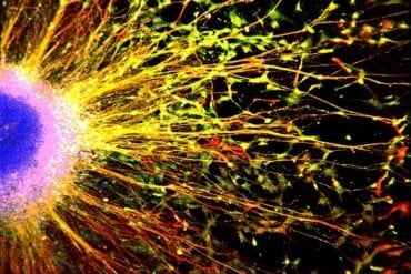
Image Source: This NeuroscienceNews.com image is credited to Harold and Margaret Milliken Hatch Laboratory of Neuroendocrinology at The Rockefeller University/Molecular Psychiatry.
Original Research: Abstract for “Stress-induced structural plasticity of medial amygdala stellate neurons and rapid prevention by a candidate antidepressant” by T Lau, B Bigio, D Zelli, B S McEwen and C Nasca in Molecular Psychiatry. Published online May 31 2016 doi:10.1038/mp.2016.68
[cbtabs][cbtab title=”MLA”]Rockefeller University. “New Signs of Stress Damage to the Brain.” NeuroscienceNews. NeuroscienceNews, 31 May 2016.
<https://neurosciencenews.com/stress-brain-damage-4347/>.[/cbtab][cbtab title=”APA”]Rockefeller University. (2016, May 31). New Signs of Stress Damage to the Brain. NeuroscienceNews. Retrieved May 31, 2016 from https://neurosciencenews.com/stress-brain-damage-4347/[/cbtab][cbtab title=”Chicago”]Rockefeller University. “New Signs of Stress Damage to the Brain.” https://neurosciencenews.com/stress-brain-damage-4347/ (accessed May 31, 2016).[/cbtab][/cbtabs]
Abstract
Stress-induced structural plasticity of medial amygdala stellate neurons and rapid prevention by a candidate antidepressant
The adult brain is capable of adapting to internal and external stressors by undergoing structural plasticity, and failure to be resilient and preserve normal structure and function is likely to contribute to depression and anxiety disorders. Although the hippocampus has provided the gateway for understanding stress effects on the brain, less is known about the amygdala, a key brain area involved in the neural circuitry of fear and anxiety. Here, in mice more vulnerable to stressors, we demonstrate structural plasticity within the medial and basolateral regions of the amygdala in response to prolonged 21-day chronic restraint stress (CRS). Three days before the end of CRS, treatment with the putative, rapidly acting antidepressant, acetyl-l-carnitine (LAC) in the drinking water opposed the direction of these changes. Behaviorally, the LAC treatment during the last part of CRS enhanced resilience, opposing the effects of CRS, as shown by an increased social interaction and reduced passive behavior in a forced swim test. Furthermore, CRS mice treated with LAC show resilience of the CRS-induced structural remodeling of medial amygdala (MeA) stellate neurons. Within the basolateral amygdala (BLA), LAC did not reduce, but slightly enhanced, the CRS-increased length and number of intersections of pyramidal neurons. No structural changes were observed in MeA bipolar neurons, BLA stellate neurons or in lateral amygdala stellate neurons. Our findings identify MeA stellate neurons as an important component in the responses to stress and LAC action and show that LAC can promote structural plasticity of the MeA. This may be useful as a model for increasing resilience to stressors in at-risk populations.
“Stress-induced structural plasticity of medial amygdala stellate neurons and rapid prevention by a candidate antidepressant” by T Lau, B Bigio, D Zelli, B S McEwen and C Nasca in Molecular Psychiatry. Published online May 31 2016 doi:10.1038/mp.2016.68


