

Summary: Researchers have discovered that the human retina synchronizes visual signals before they even reach the brain. By adjusting the diameter of nerve fibers, longer retinal axons transmit signals faster, ensuring they arrive in near-perfect sync with shorter ones.
This process reduces timing differences to just milliseconds, preserving a unified visual experience. The finding challenges the long-held assumption that such synchronization occurs only in the brain.
Key Facts:
- Built-In Synchronization: Longer retinal nerve fibers are thicker, enabling faster conduction to match shorter pathways.
- Millisecond Precision: Timing differences are reduced to just a few milliseconds.
- Beyond the Brain: Visual timing coordination starts in the retina, not solely in the brain.
Source: IOB
Perception depends not only on what we see, but also on when we see it.
Signals initiated by light-sensitive cells in the retina travel through nerve fibers of varying lengths before converging at the optic nerve and continuing to the brain.
Even neighboring cells in the central retina can transmit signals over very different distances – raising the question of how the brain avoids receiving a scrambled or delayed picture of the world.
A study published by researchers of the Institute of Molecular and Clinical Ophthalmology Basel (IOB) in Nature Neuroscience shows that differences in the speed and distance of nerve signals are actively balanced within the human eye itself to support a unified and temporally accurate visual experience.
Key findings:
- Axonal tuning in the human retina: Longer axons of retinal ganglion cells have larger diameters and faster conduction speeds, which helps align signal arrival times.
- Precision in milliseconds: This mechanism helps align the timing of signals from different parts of the retina, reducing differences in arrival time to just a few milliseconds.
- Multiple layers of compensation: Alongside axon speed, other factors like the initial response time of retinal cells and further adjustments in the brain contribute to synchronization.
IOB researchers reveal that the fine-tuning of visual timing in humans begins not in the brain but in the retina, thus challenging previous assumptions.
They show that this built-in compensation helps maintain the clarity and consistency of what we see – despite structural differences in how signals are routed through the eye.
These findings raise important questions about how nerve fibers are adjusted during development – for example, how their diameter is regulated and how their membranes help control the speed of signal transmission.
Understanding these mechanisms could reveal fundamental principles of temporal coordination in the brain, with relevance well beyond the visual system.
About this visual neuroscience research news
Author: Elsa Sigle
Source: IOB
Contact: Elsa Sigle – IOB
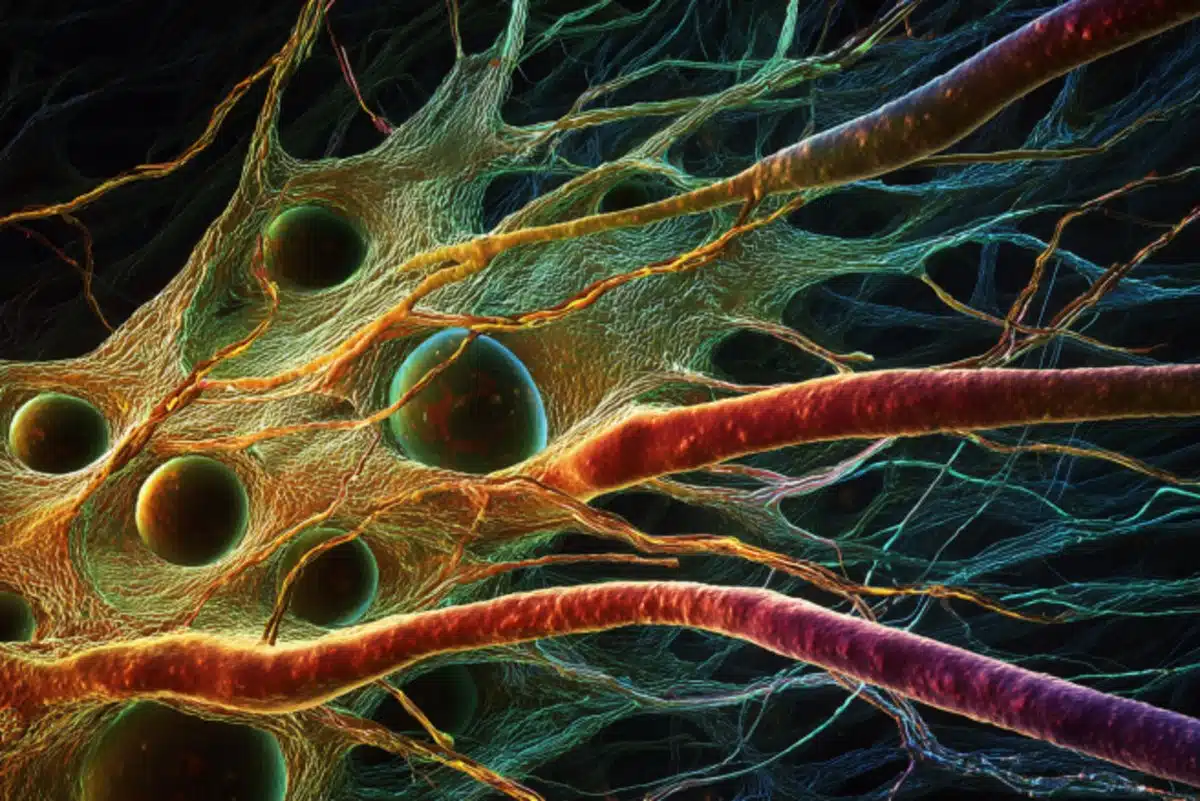
Image: The image is credited to Neuroscience News
Original Research: Open access.
“Synchronization of visual perception within the human fovea” by Felix Franke et al. Nature Neuroscience
Abstract
Synchronization of visual perception within the human fovea
The human brain constructs a model of the world by processing sensory signals with distinct temporal characteristics that may differ in generation and transmission speed within a single sensory modality.
To perceive simultaneous events as occurring at the same time, the brain must synchronize this sensory information, yet the mechanisms underlying such synchronization remain unclear.
By combining human neural recordings, behavioral measurements and modeling, we show that in the human visual system, this process begins in the fovea centralis, the retinal region used for reading and recognizing faces.
Reaction times to foveal single-cone photostimulation were similar across the central visual field, although visual information from neighboring foveal cones travels along axons of highly different lengths.
From direct measurements of action potential propagation speeds, axon diameters and lengths in the human fovea centralis, we found that longer foveal axons have larger diameters and increased propagation speeds.
We conclude that the human brain orchestrates axonal conduction speeds of unmyelinated axons in the retina to synchronize the arrival times of sensory signals.
These results suggest a previously unknown mechanism by which the human brain synchronizes perception.


