Summary: No two brains are shaped exactly alike, yet most neural implants use a “one-size-fits-all” design. Researchers have developed a breakthrough approach to 3D printing soft, stretchable bioelectrodes tailored to the specific ridges (gyri) and grooves (sulci) of an individual’s brain.
These hydrogel-based sensors provide nearly perfect connectivity and better signal quality without damaging sensitive brain tissue or disrupting fluid transport.
Key Findings
- Superior Signal: Because the electrodes follow the brain’s unique structure precisely, they maintain “nearly perfect” connectivity, resulting in higher-quality data for monitoring diseases.
- Biocompatibility: In rat models, the sensors remained effective for 28 days with zero immune response, proving they are safe for long-term “implantation.”
- Pizza-Sized Complexity: The researchers noted that if an adult brain were spread flat, it would cover 2,000 square centimeters (the size of two large pizzas). Their 3D-printed mesh is the first to navigate this vast, folded terrain comfortably.
- Commercial Scalability: This framework provides a roadmap for mass-producing patient-specific bioelectrodes for both monitoring and potentially treating neurodegenerative disorders.
Source: Penn State
Soft electrodes designed to perfectly match a person’s brain surface may help advance neural interfaces for neurodegenerative disease monitoring and treatment, according to a new study led by Penn State researchers.
Neural interfaces are powered by tiny sensors capable of tracking biophysical signals, known as bioelectrodes. These sensors are usually made from stiff materials in a one-size-fits-all design that struggles to match the brain’s complex structure.
The researchers have created a novel approach to 3D printing bioelectrodes that can stretch and morph to fit the minor differences that make every brain unique.

The team used software to simulate detailed brains based on MRI scans taken from 21 human patients, shaping a set of electrodes tailored for brains’ specific structures before 3D printing the electrodes and models of the brains.
In a paper published in Advanced Materials, they reported that their electrodes better fit the structure of the brain than traditional designs, while remaining effective and biologically compatible, even in tests done in rats.
The folds in the human brain are created through a process known as gyrification, where the cortical sheet on the outer wall of the brain bunches up into ridges, known as gyri, and grooves, known as sulci. This helps cells across the brain communicate at high speeds, and allows for a relatively large organ to fit compactly in the skull — a spread-out adult brain would be around 2,000 square centimeters, or about the size of two large pizzas.
Although the major cortical folds are consistent across individuals, the precise layout of the brain’s gryi and sulci changes substantially from person to person, according to Tao Zhou, Wormley Family Early Career Professor, assistant professor of engineering science and mechanics and corresponding author on the paper. However, traditional bioelectrode designs don’t take this into account.
“Each person has a different brain structure, depending on their height, weight, age, sex and more,” said Zhou, who also holds an affiliation in biomedical engineering and the center for neural engineering at Penn State.
“Despite this, we try to fit neural interfaces onto brains like they have identical structures. This motivated us to create electrodes that are tailored for each individual, based on the structure of their brain.”
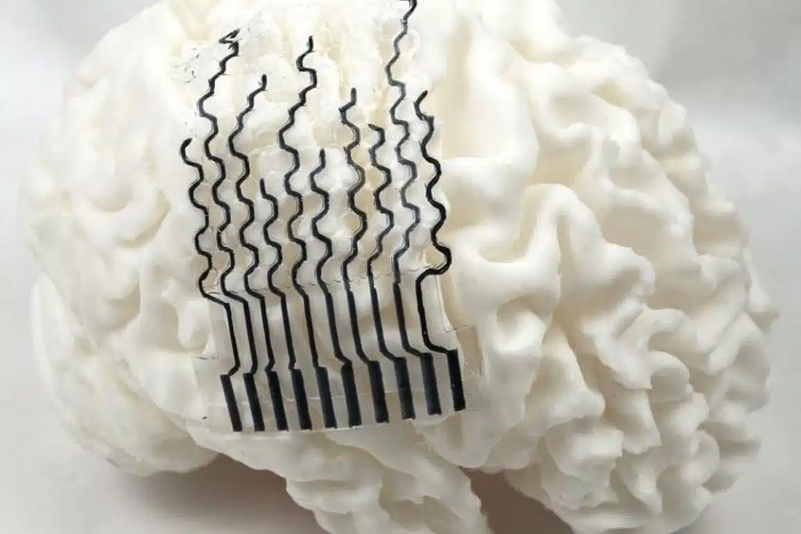
The electrodes are built mainly from a water-rich material known as hydrogel to better match with the soft tissues and patient-specific geometry of a brain. Furthermore, the team used a novel honeycomb-inspired structure that offers flexibility and strength, while remaining cost-effective and quick to print, according to Zhou.
“The honeycomb structure helps us significantly reduce the stiffness of the electrodes, without sacrificing their mechanical strength,” Zhou said. “What’s more, the structure helps us reduce the overall material used during fabrication, reducing production time, cost and environmental impact.”
Production starts by taking an MRI scan of a patient’s brain, which is used to conduct finite element analysis — a process that creates a detailed simulation of a person’s neural structure. This analysis is then rendered as a 3D model of the patient’s brain, where the team uses computer software to tailor a bioelectrode specifically morphed to fit the ridges and grooves of the cerebral cortex.
After shaping, the team 3D prints the hydrogel electrode using direct ink printing, a technique that can create electrodes capable of monitoring and transmitting brain signals over a relatively small surface. For this study, the team 3D printed models of 21 different participant brains, applying their electrodes and physically measuring how accurately the electrodes could fit the brain surface.
Zhou explained how traditional fabrication approaches require specialized facilities like clean rooms, making them incredibly expensive to customize — 3D printing allows the team to personalize and manufacture electrodes much faster, for a fraction of the price.
Compared to traditional approaches, the hydrogel-based electrodes follow the structure of the brain more precisely. Zhou said their approach produces electrodes that exhibit nearly perfect connectivity to electrical signals present in the brain. Additionally, because the stretchy gel is so malleable, it can be applied to the soft brain tissue without causing damage, compared to the stiff materials comprising other designs that could damage tissue.
According to Zhou, the softness of their electrodes enables closer and more stable contact with the brain, in turn facilitating higher-quality, more reliable monitoring. Moreover, bioelectrodes made with this approach don’t impact fluid transport around the brain, a critical aspect of brain function that many traditional electrodes disrupt.
“Personalizing the electrodes to the brain’s specific structure substantially improves their reliability,” Zhou said. “Because they conform to the brain better, the signal quality itself is significantly improved.”
To further study their electrodes, the team placed them onto the brains of rat models over a period of 28 days. The rats did not exhibit any immune response to the printed electrodes, a key consideration in biodevice development, Zhou said. Additionally, the electrodes did not exhibit performance degradation, while offering sensitive and accurate readings of the electric and physiological signals in the brain.
Zhou said he believes that this printing method could serve as a framework for the commercial-scale printing of bioelectrodes customized for specific patients. Although these systems are traditionally used for monitoring neural activity, the team plans to explore how personalized electrodes may contribute to neurological treatments.
“We are looking to further improve this technology to optimize the electrodes to monitor for specific diseases,” Zhou said. “In the future, we would really like to work with patients to see how this approach could support brain monitoring and disease treatment in clinical settings.”
Additional co-authors affiliated with Penn State include Nanyin Zhang, professor of biomedical engineering and Dorothy Foehr Huck and J. Lloyd Huck Chair in Brain Imaging; Sulin Zhang, professor of engineering science and mechanics and of biomedical engineering; engineering science and mechanics doctoral candidates Marzia Momin, Luyi Feng, Salahuddin Ahmed and Jiashu Ren; biomedical engineering doctoral candidates Xiaoai Chen, Hyunjin Lee and post-doctoral scholar Samuel R. Cramer; mechanical engineering doctoral candidate Xinyi Wang; Basma AlMahood, an undergraduate student studying physics at the time of research who is now a physics doctoral candidate at Michigan State University; and Li-Pang Huang, a research assistant.
Funding: This work was supported by the U.S. National Science Foundation and the National Institutes of Health.
Key Questions Answered:
A: They are made primarily of hydrogel, a material that is mostly water. This allows them to “morph” and stretch along with the brain’s natural movements, making them feel more like a part of the organ rather than a foreign object.
A: While the current focus is on medical treatment for diseases like Parkinson’s or epilepsy, the ability to 3D print custom-fit sensors quickly and cheaply definitely paves the way for more comfortable consumer neural interfaces in the future.
A: Nature knows best! The honeycomb structure provides maximum strength with minimum material. It makes the electrode sturdy enough to handle but flexible enough to sink into the deep “grooves” (sulci) of the brain without snapping.
Editorial Notes:
- This article was edited by a Neuroscience News editor.
- Journal paper reviewed in full.
- Additional context added by our staff.
About this neurotech research news
Author: Ty Tkacik
Source: Penn State
Contact: Ty Tkacik – Penn State
Image: The image is credited to Tao Zhou
Original Research: Open access.
“3D-Printable, Honeycomb-Inspired Tissue-Like Bioelectrodes for Patient-Specific Neural Interface” by Marzia Momin, Luyi Feng, Xiaoai Chen, Salahuddin Ahmed, Basma AlMahmood, Li-Pang Huang, Jiashu Ren, Xinyi Wang, Hyunjin Lee, Samuel R. Cramer, Nanyin Zhang, Sulin Zhang, Tao Zhou. Advanced Materials
DOI:10.1002/adma.202516291
Abstract
3D-Printable, Honeycomb-Inspired Tissue-Like Bioelectrodes for Patient-Specific Neural Interface
The unique gyral patterns of the human brain demand patient-specific neural interfaces to achieve precise neuromodulation, mitigate adverse tissue responses, and optimize therapeutic efficacy and safety.
One-size-fits-all, conventional rigid electrocorticography (ECoG) electrodes, standardized for mass production through lithographic techniques, exhibit limited conformability to the brain’s heterogeneous cortical topography.
This mechanical mismatch results in poor electrode-tissue contact, signal loss, and foreign body responses.
To address these limitations, we present an integrated novel platform, synergizing MRI-based anatomical mapping, finite element analysis (FEA)—optimized mechanical design, and direct ink writing (DIW) 3D printing to fabricate electrodes customized to individual gyral patterns.
The resulting honeycomb-inspired printable gel electrode (HiPGE) employs a bioinspired honeycomb architecture with ultra-soft hydrogels, engineered to match the bending stiffness of brain tissue (0.1–10 kPa) while maintaining cost-efficiency and long-term durability.
This mechanical congruence ensures exceptional cortical conformability and adaptive interfacing, circumventing the geometric and material limitations of traditional rigid electrodes.
By combining patient-specific design with scalable fabrication, our platform establishes a transformative framework for neural interface engineering, enhancing precision, biocompatibility, and functional performance in neuromodulation therapies and neuroprosthetic applications.