Summary: Researchers revealed the intricate role of OPC cells in shaping the brain’s neural circuits. Previously known mainly for supporting neuron insulation, OPCs also prune unnecessary synaptic connections, refining the brain’s wiring as we accumulate experiences.
A new imaging technique has enabled researchers to track OPCs as they selectively prune synapses, helping to maintain healthy brain function. The findings could inform research on complex brain diseases, such as glioma and Alzheimer’s, as OPCs are now suspected of playing a role in these conditions. By mapping OPC activity in unprecedented detail, researchers are paving the way to better understand the brain’s adaptive processes.
Key Facts
- OPCs are responsible for pruning unnecessary synaptic connections in the brain.
- New imaging techniques allow scientists to observe OPC activity at an unprecedented scale.
- OPC cells, once thought to only support neurons, may also influence brain disease progression.
Source: CSHL
Imagine yourself sometime in the far future aboard a routine rocket to Mars. Someone just spilled their drink. Without gravity, it collects in floating blobs that ripple right before your eyes. Now freeze.
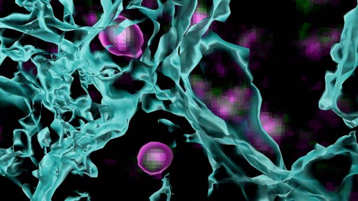
What you see might look something like the above image from Cold Spring Harbor Laboratory’s (CSHL’s) Cheadle lab. But those purple and green blobs aren’t the floating remains of somebody’s drink. They’re mysterious cells in the brain’s visual cortex called OPCs.

The visual cortex processes everything we see. Incoming visual information is relayed to this outer layer of the brain via synapses—the silver streaks above.
When the brain’s neural circuits are first wired up, more connections, or synapses, are created than needed. As the brain accumulates new experiences and information, OPCs shape neural circuitry by pruning unnecessary synapses.
“OPCs are doing all sorts of things in the brain that help it to function in a normal, healthy way,” CSHL Assistant Professor Lucas Cheadle says. OPCs are a specialty of the Cheadle lab. He and his team discovered OPCs’ function as neural landscapers in 2022.
Before that, they were thought only to produce oligodendrocytes, cells that sheath and support neurons. Now, Cheadle has developed new ways to zoom in and see OPCs in action.
“We’re able to see what thousands of OPCs, and even smaller groups of 30-50, are doing,” he explains.
“From there, we can figure out which synapses are fully engulfed by an OPC, which are in the process of being pruned, and which have maybe just been checked on by an OPC but not processed.”
The new techniques used to produce the image above have become essential tools in Cheadle’s ongoing work. He and his team are now building on their 2022 discovery to help paint a complete picture of OPCs’ role in health and disease.
Cheadle explains: “These mysterious cells are one of the primary sources of glioma,” a deadly brain cancer. “They’re potentially involved in Alzheimer’s disease as well.”
It’ll take more research to illustrate these connections in detail. In the meantime, Cheadle is eager to share his lab’s new tools with researchers around the world.
“The brain is constantly changing, and the same approaches you’d use to look at one type of cell can’t just be applied across the board,” he says. “We’re adapting and innovating to keep up with it—to better understand how the brain works.”
About this visual neuroscience research news
Author: Sara Giarnieri
Source: CSHL
Contact: Sara Giarnieri – CSHL
Image: The image is credited to Cheadle lab/Cold Spring Harbor Laboratory
Original Research: Open access.
“High-confidence and high-throughput quantification of synapse engulfment by oligodendrocyte precursor cells” by Lucas Cheadle et al. Nature Protocols
Abstract
High-confidence and high-throughput quantification of synapse engulfment by oligodendrocyte precursor cells
Oligodendrocyte precursor cells (OPCs) sculpt neural circuits through the phagocytic engulfment of synapses during development and adulthood. However, existing techniques for analyzing synapse engulfment by OPCs have limited accuracy.
Here we describe the quantification of synapse engulfment by OPCs via a two-pronged cell biological approach that combines high-confidence and high-throughput methodologies.
Firstly, an adeno-associated virus encoding a pH-sensitive, fluorescently tagged synaptic marker is expressed in neurons in vivo to differentially label presynaptic inputs, depending upon whether they are outside of or within acidic phagolysosomal compartments.
When paired with immunostaining for OPC markers in lightly fixed tissue, this approach quantifies the engulfment of synapses by around 30–50 OPCs in each experiment.
The second method uses OPCs isolated from dissociated brain tissue that are then fixed, incubated with fluorescent antibodies against presynaptic proteins, and analyzed by flow cytometry, enabling the quantification of presynaptic material within tens of thousands of OPCs in <1 week.
The integration of both methods extends the current imaging-based assays, originally designed to quantify synaptic phagocytosis by other brain cells such as microglia and astrocytes, by enabling the quantification of synaptic engulfment by OPCs at individual and populational levels.
With minor modifications, these approaches can be adapted to study synaptic phagocytosis by numerous glial cell types in the brain. The protocol is suitable for users with expertise in both confocal microscopy and flow cytometry.
The imaging-based and flow cytometry-based protocols require 5 weeks and 2 d to complete, respectively.


