Summary: A novel non-invasive method merges acoustic imaging methods with new algorithms to explore changes in white and gray matter following head injury.
Source: Acoustical Society of America
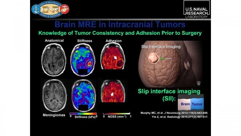
A noninvasive method to measure the stiffness parameters along fibrous pathways within the brain is helping researchers explore traumatic brain injuries. The stiffness of these tissues can reveal clues about changes and pathologies within the brain’s gray and white matter.
During the 179th Meeting of the Acoustical Society of America, which will be held virtually Dec. 7-10, Anthony J. Romano, of the U.S. Naval Research Laboratory, will describe the method known as waveguide elastography. His session, “An overview of mixed-model inversion and its application to the study of traumatic brain injury,” will be held Dec. 9 at noon Eastern U.S.
Romano went from working on antisubmarine warfare, analyzing acoustic signatures during the Cold War, to converting his submarine-detection algorithms to nondestructive evaluation for biomedical applications.
Waveguide elastography merges magnetic resonance elastography (MRE) and diffusion tensor imaging (DTI) with a combination of isotropic and anisotropic inversion algorithms.
“It was an ‘aha!’ moment when I realized I could use these technologies with my algorithms to explore both gray and white matter inside the brain,” said Romano.
An MRE provides dynamic elastic displacement by vibrating the skull and using equations of motion. This displacement isn’t the same as stiffness but is related to stiffness through equations of motion.
Romano and colleagues are studying the University of Delaware hockey team’s brains. They scanned players’ brains pre-, mid-, and post-season and found significant increases in stiffness as the season progressed because of concussions every player experienced.

“Stiffness is a much more reliable metric than displacement due to SNR, so this method allows us to evaluate stiffness from the measured strain waves within tissues for diagnostic purposes,” said Romano.
DTI simply measures the diffusion of water along fibrous pathways, such as myelinated axons within the brain. The brain’s white matter consists mainly of myelinated axons, which form fibrous structures that can act as waveguides for the anisotropic propagation of sound.
“Waves can propagate at different speeds and stiffnesses in the direction of propagation,” Romano said.
Gray matter “is isotropic, which means the stiffness is the same in every direction of sound propagation — unless there is a stiffer or softer inclusion,” he said.
Applying algorithms in this situation helps reveal brain health. Gray matter gets an isotropic shear wave equation, while white matter gets an orthotropic inversion algorithm.
About this TBI research news
Source: Acoustical Society of America
Contact: Press Office – Acoustical Society of America
Image: The image is credited to Anthony J. Romano
Original Research: The findings will be presented at 179th Meeting of the Acoustical Society of America


