

These results are the first step in a project to push back the limits of existing dMRI imaging technology, to improve diagnosis and investigate potential treatments for brain diseases.
The first analysis of biological processes within brain tissue using neutrons at the Institut Laue-Langevin has revealed that the common application of formaldehyde preservatives changes, rather than maintains, fundamental properties such as rates of water diffusion. The mapping of cellular water in the brain is a key factor in the post-mortem analysis of several brain pathologies (including tumours and multiple sclerosis), with a view to earlier diagnosis and potential treatment. These results suggest the need for a review of existing research in this area.
The results are the first stage in the team’s own pioneering application of neutrons to understand in unprecedented detail the movement of cellular water within brain tissue. The analysis of this movement is generally performed by diffusion magnetic resonance imaging (dMRI), and provides the basis for diagnosing several brain diseases. These first results clearly demonstrate neutrons’ ability to ‘see’ the effects of these biological processes on a scale 10,000 times smaller than dMRI. In future ILL’s neutrons will analyse with unprecedented resolution cellular water dynamics in ex-vivo pathology-bearing brain tissue samples, thus helping doctors spot the early signs of these diseases and investigate potential treatments.

Cellular water is the major constituent of our body and its content may vary in brain regions depending on their specific composition. Water plays a key role in cell regulation, and its distribution and movement is an accurate indicator of cellular structure; this is because it interacts with different tissue components such as membranes and nerve fibres.
dMRI and other imaging techniques use water diffusion as a contrast method for revealing and characterising several brain pathologies (i.e. ischemia, tumours and, recently, inherited prion disease) on the micron scale 100 times thinner than a human hair. At this scale, however, the contribution of the macromolecular components cannot be separated and have to be averaged out instead.
As the standard imaging techniques used to detect the early signs of brain pathologies are limited in resolution, the use of preservation techniques to investigate pathological conditions in ex-vivo specimens has increased. However, there are concerns over the impact of these preservation processes on our tissues’ fundamental structural and compositional properties, and this has undermined confidence in this line of research.
To address these concerns Dr Francesca Natali from the Italian CNR (Consiglio Nazionale delle Ricerche), in collaboration with Dr Yuri Gerelli, a scientist from the Institut Laue-Langevin, the world’s flagship centre for neutron science, Prof. J. Peters from France’s Joseph Fourier University in Grenoble (UJF), and Dr Calogero Stelletta from the University of Padova in Italy compared the behaviour of cellular water in ex-vivo bovine tissue preserved using two common preservation techniques: chemical fixation, using formaldehyde solutions, and cryo-preservation, where cells or whole tissues are cooled to sub-zero temperatures.
The researchers obtained fresh post-mortem bovine brains from an Italian slaughter-house and applied the different preservation techniques. These samples were then investigated using incoherent quasi-elastic neutron scattering (QENS) on the high-resolution IN5 spectrometer at the Institut Laue-Langevin (ILL).
Neutrons are an ideal probe for the investigation of biological materials at the atomic scale. As they produce no damaging radiation effects, they can accurately map any change in the samples over time.
From this analysis Dr Francesca Natali and her colleagues identified a significant reduction of water movement as a result of the introduction of the formaldehyde-based preservation solutions (potentially due to the formation of cross-links between proteins, within which free water may become trapped, reducing its mobility). This effect was not seen in the samples that underwent cryo-preservation.
As well as these findings, the results of this study also demonstrate for the first time the power of neutrons to model cellular water diffusion within brain tissue; this new modeling technique could help dMRI specialists push back the limits of existing imaging technology, to improve their diagnoses and investigate potential treatments for brain pathologies.
In a separate study, the same team are investigating how the movement and distribution of cellular water in brain tissue is affected by myelin, an electrical insulator that forms protective layers known as sheaths around brain cell axons. Myelin is responsible for speeding up electrical impulses as they travel along tissue fibres. Many neurodegenerative autoimmune diseases, including multiple sclerosis, are caused by the degradation of myelin over time. The neutron scattering team’s new understanding of the impact on research results of preservation techniques will enhance its atomic-scale investigations into the conditions underlying autoimmune diseases and the potential for treatment.
Notes about this neuroscience and neuroimaging research
Contact: Mr James Romero – Institut Laue-Langevin
Source: Institut Laue-Langevin press release
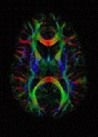
Image Source: the bovine brain tissue exposed to a neutron beam image is adapted from the ILL press release. The dMRI brain scan image is available in the public domain.
Original Research: Abstract for “Anomalous proton dynamics of water molecules in neural tissue as seen by quasi-elastic neutron scattering. Impact on medical imaging techniques” by F. Natali, Y. Gerelli, C. Stelletta, and J. Peters in AIP Conference Proceedings. Published online December 2012 doi: 10.1063/1.4794632


