

Summary: Researchers team up with special effects experts to create a life-like 3D simulator that allows surgeons to practice delicate brain surgeries.
Source: Johns Hopkins Medicine.
Special effects pros help create lifelike 3-D simulator for practicing brain surgery.
A team of computer engineers and neurosurgeons, with an assist from Hollywood special effects experts, reports successful early tests of a novel, lifelike 3D simulator designed to teach surgeons to perform a delicate, minimally invasive brain operation.
A report on the simulator that guides trainees through an endoscopic third ventriculostomy (ETV) was published in the Journal of Neurosurgery: Pediatrics on April 25. The procedure uses endoscopes, which are small, computer-guided tubes and instruments, to treat certain forms of hydrocephalus, a condition marked by an excessive accumulation of cerebrospinal fluid and pressure on the brain. ETV is a minimally invasive procedure that short-circuits the fluid back into normal channels in the brain, eliminating the need for implantation of a shunt, a lifelong device with the associated complications of a foreign body.
“For surgeons, the ability to practice a procedure is essential for accurate and safe performance of the procedure. Surgical simulation is akin to a golfer taking a practice swing,” says Alan R. Cohen, M.D., professor of neurosurgery at the Johns Hopkins University School of Medicine and a senior author of the report. “With surgical simulation, we can practice the operation before performing it live.”
While cadavers are the traditional choice for such surgical training, Cohen says they are scarce, expensive, nonreusable, and most importantly, unable to precisely simulate the experience of operating on the problem at hand, which Cohen says requires a special type of hand-eye coordination he dubs “Nintendo Neurosurgery.”
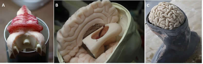
In an effort to create a more reliable, realistic and cost-effective way for surgeons to practice ETV, the research team worked with 3D printing and special effects professionals to create a lifelike, anatomically correct, full-size head and brain with the touch and feel of human skull and brain tissue.
The fusion of 3D printing and special effects resulted in a full-scale reproduction of a 14-year-old child’s head, modeled after a real patient with hydrocephalus, one of the most common problems seen in the field of pediatric neurosurgery. Special features include an electronic pump to reproduce flowing cerebrospinal fluid and brain pulsations. One version of the simulator is so realistic that it has facial features, hair, eyelashes and eyebrows.
To test the model, Cohen and his team randomly paired four neurosurgery fellows and 13 medical residents to perform ETV on either the ultra-realistic simulator or a lower-resolution simulator, which had no hair, lashes or brows.
After completing the simulation, fellows and residents each rated the simulator using a five-point scale. On average, both the surgical fellows and the residents rated the simulator more highly (4.88 out of 5) on its effectiveness for ETV training than on its aesthetic features (4.69). The procedures performed by the trainees were also recorded and later watched and graded by two fully trained neurosurgeons in a way that they could not identify who the trainees were or at what stage they were in their training.
The neurosurgeons assessed the trainees’ performance using criteria such as “flow of operation,” “instrument handling” and “time and motion.”
Neurosurgeons consistently rated the fellows higher than residents on all criteria measured, which accurately reflected their advanced training and knowledge, and demonstrated the simulator’s ability to distinguish between novice and expert surgeons.
Cohen says that further tests are needed to determine whether the simulator will actually improve performance in the operating room. “With this unique assortment of investigators, we were able to develop a high-fidelity simulator for minimally invasive neurosurgery that is realistic, reliable, reusable and cost-effective. The models can be designed to be patient-specific, enabling the surgeon to practice the operation before going into the operating room,” says Cohen.
Other authors on this paper include Roberta Rehder from the Johns Hopkins School of Medicine, and Peter Weinstock, Sanjay P. Parbhu, Peter W. Forbes and Christopher Roussin from Boston Children’s Hospital.
Funding: Funding for the study was provided by a grant from the Boston Investment Conference. The research team acknowledges the contribution of FracturedFX, an Emmy Award-winning special effects group from Hollywood, California, in the development of the surgical models.
The investigators report no financial stake or interests in the success of the simulator.
Source: Chanapa Tantibanchachai – Johns Hopkins Medicine
Image Source: NeuroscienceNews.com image is credited to AANS.
Original Research: Full open access research for “Creation of a novel simulator for minimally invasive neurosurgery: fusion of 3D printing and special effects” by Peter Weinstock, MD, PhD, Roberta Rehder, MD, Sanjay P. Prabhu, MBBS, FRCR, Peter W. Forbes, PhD, Christopher J. Roussin, PhD, and Alan R. Cohen, MD in Journal of Neurosurgery: Pediatrics. Published online April 25 2017 doi:10.3171/2017.1.PEDS16568
[cbtabs][cbtab title=”MLA”]Johns Hopkins Medicine “When Hollywood Met Neurosurgery.” NeuroscienceNews. NeuroscienceNews, 25 April 2017.
<https://neurosciencenews.com/neurosurgery-special-effects-props-6497/>.[/cbtab][cbtab title=”APA”]Johns Hopkins Medicine (2017, April 25). When Hollywood Met Neurosurgery. NeuroscienceNew. Retrieved April 25, 2017 from https://neurosciencenews.com/neurosurgery-special-effects-props-6497/[/cbtab][cbtab title=”Chicago”]Johns Hopkins Medicine “When Hollywood Met Neurosurgery.” https://neurosciencenews.com/neurosurgery-special-effects-props-6497/ (accessed April 25, 2017).[/cbtab][/cbtabs]
Abstract
Creation of a novel simulator for minimally invasive neurosurgery: fusion of 3D printing and special effects
OBJECTIVE
Recent advances in optics and miniaturization have enabled the development of a growing number of minimally invasive procedures, yet innovative training methods for the use of these techniques remain lacking. Conventional teaching models, including cadavers and physical trainers as well as virtual reality platforms, are often expensive and ineffective. Newly developed 3D printing technologies can recreate patient-specific anatomy, but the stiffness of the materials limits fidelity to real-life surgical situations. Hollywood special effects techniques can create ultrarealistic features, including lifelike tactile properties, to enhance accuracy and effectiveness of the surgical models. The authors created a highly realistic model of a pediatric patient with hydrocephalus via a unique combination of 3D printing and special effects techniques and validated the use of this model in training neurosurgery fellows and residents to perform endoscopic third ventriculostomy (ETV), an effective minimally invasive method increasingly used in treating hydrocephalus.
METHODS
A full-scale reproduction of the head of a 14-year-old adolescent patient with hydrocephalus, including external physical details and internal neuroanatomy, was developed via a unique collaboration of neurosurgeons, simulation engineers, and a group of special effects experts. The model contains “plug-and-play” replaceable components for repetitive practice. The appearance of the training model (face validity) and the reproducibility of the ETV training procedure (content validity) were assessed by neurosurgery fellows and residents of different experience levels based on a 14-item Likert-like questionnaire. The usefulness of the training model for evaluating the performance of the trainees at different levels of experience (construct validity) was measured by blinded observers using the Objective Structured Assessment of Technical Skills (OSATS) scale for the performance of ETV.
RESULTS
A combination of 3D printing technology and casting processes led to the creation of realistic surgical models that include high-fidelity reproductions of the anatomical features of hydrocephalus and allow for the performance of ETV for training purposes. The models reproduced the pulsations of the basilar artery, ventricles, and cerebrospinal fluid (CSF), thus simulating the experience of performing ETV on an actual patient. The results of the 14-item questionnaire showed limited variability among participants’ scores, and the neurosurgery fellows and residents gave the models consistently high ratings for face and content validity. The mean score for the content validity questions (4.88) was higher than the mean score for face validity (4.69) (p = 0.03). On construct validity scores, the blinded observers rated performance of fellows significantly higher than that of residents, indicating that the model provided a means to distinguish between novice and expert surgical skills.
CONCLUSIONS
A plug-and-play lifelike ETV training model was developed through a combination of 3D printing and special effects techniques, providing both anatomical and haptic accuracy. Such simulators offer opportunities to accelerate the development of expertise with respect to new and novel procedures as well as iterate new surgical approaches and innovations, thus allowing novice neurosurgeons to gain valuable experience in surgical techniques without exposing patients to risk of harm.
“Creation of a novel simulator for minimally invasive neurosurgery: fusion of 3D printing and special effects” by Peter Weinstock, MD, PhD, Roberta Rehder, MD, Sanjay P. Prabhu, MBBS, FRCR, Peter W. Forbes, PhD, Christopher J. Roussin, PhD, and Alan R. Cohen, MD in Journal of Neurosurgery: Pediatrics. Published online April 25 2017 doi:10.3171/2017.1.PEDS16568


