

Summary: Scientists discovered how the brain uses objects to anchor our sense of direction, solving part of the mystery of spatial navigation. Experiments in mice showed that cells in the postsubiculum fired strongly when facing an object, while cells for other directions were suppressed, sharpening orientation.
This mechanism suggests that object recognition is tightly integrated with spatial navigation. The findings could explain why people with Alzheimer’s and related conditions often lose their sense of place and direction.
Key Facts
- Object Anchors: Postsubiculum cells fire in response to objects, reinforcing direction sense.
- Dual Systems: Visual and navigation systems interact to guide orientation.
- Disease Link: Early Alzheimer’s damages orientation regions, leading to disorientation.
Source: McGill University
We take our understanding of where we are for granted, until we lose it. When we get lost in nature or a new city, our eyes and brains kick into gear, seeking familiar objects that tell us where we are.
How our brains distinguish objects from background when finding direction, however, was largely a mystery. A new study provides valuable insight into this process, with possible implications for disorientation-causing conditions such as Alzheimer’s.
The scientists, based at The Neuro (Montreal Neurological Institute-Hospital) of McGill University and the University Medical Center Göttingen, ran an experiment with mice using ultrasound imaging to measure and record brain activity. The mice were shown visual stimuli, either an object or a scrambled image showing no distinct object.
They found a small number of brain areas that fired especially when the mouse looked at objects. These areas were found in a brain region called the postsubiculum which specializes in keeping track of where the animal is facing at any given time. Each direction activates a specific cell in the postsubiculum.
Objects in the mice’s vision increased the firing of the cell responsible for the direction in which the mouse was looking. They also inhibited cells responsible for directions where the mouse was not looking. Together, this activity reinforced the mouse’s perception of where it was relative to the object.
While the postsubiculum was particularly sensitive to the presence of objects in the mouse’s vision, other brain regions were not, suggesting that object recognition is particularly important to the brain’s understanding of where it is and where the animal is looking.
This finding offers clues as to why humans with diseases such as dementia and Alzheimer’s often lose track of where they are. A recent study from Oxford University has shown that the accumulation of tau protein-a hallmark of Alzheimer’s-happens first in the brain regions responsible for spatial orientation.
“A very useful aspect of our study is it presents a very high-level understanding of two systems that interact together-the visual and spatial recognition systems,” says Stuart Trenholm, a researcher at The Neuro and the paper’s co-senior author.
“We have a decent understanding now of how they modulate each other. They are both very high-level brain functions and lot of these neurodegenerative disorders lead to disconnections between these states, so that will be interesting to look into in the future.”
“Our results are incredibly surprising,” says Adrien Peyrache, a researcher at The Neuro and the paper’s co-senior author.
“Nobody would have predicted that object processing would occur in the navigation system and not in the visual cortex. For the first time, we have an inside-the-brain perspective of what an object is, and how we use an object to get a sense of the world around us.”
About this neuroscience and navigation research news
Author: Shawn Hayward
Source: McGill University
Contact: Shawn Hayward – McGill University
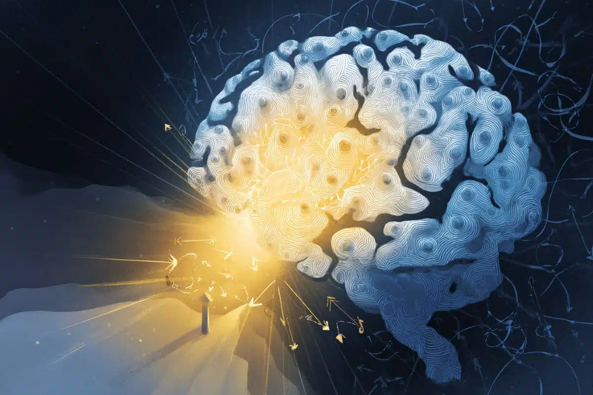
Image: The image is credited to Neuroscience News
Original Research: Closed access.
“Visual objects refine head direction coding” by Adrien Peyrache et al. Science
Abstract
Visual objects refine head direction coding
INTRODUCTION
In our day-to-day life, we use visual objects (e.g., a specific clock tower when walking around town, etc.) as spatial landmarks to help orientate ourselves as we make our way through the world. We can use visual objects as spatial landmarks because our brains dedicate considerable computational power toward parsing the world into objects.
Moreover, our location in the world is processed by specialized cells such as place cells, grid cells, and head direction (HD) cells in the brain’s spatial navigation system. However, much remains unknown about how visual objects modulate tuning properties of the neurons that encode spatial variables, partly because it is unclear how visual objects are processed in the rodent brain, where spatial navigation signals have been most extensively studied.
RATIONALE
To address this gap in knowledge, we reasoned that a brainwide activity screen in mice could identify visual object–preferring areas in an unbiased way. Using electrophysiology, we then characterized the spatial correlates of individual neurons in the areas responding to objects in freely moving mice.
RESULTS
Our brainwide screen based on functional ultrasound imaging revealed that spatial navigation–related areas, not visual cortical areas, tended to show a preference for visual objects. The postsubiculum, a hub of the HD system, exhibited the strongest object preference. In head-fixed conditions, we discovered that HD cells were differentially modulated by visual stimulation based on their preferred firing direction: HD cells pointing toward the visual stimulus were excited, whereas HD cells pointing away from the visual stimulus were inhibited. The same modulation was observed in freely moving conditions, when an object was displayed in the environment.
CONCLUSION
Visual objects refine the population encoding of HD in the postsubiculum by increasing the response of HD cells that code toward the object while simultaneously decreasing the firing rate of HD cells that code directions away from the object. As an analogy, imagine yourself wandering around a city with a compass (which, in our experiments, is the brain’s HD system), and each time you look at a landmark (which, in our experiments, is an image of an object), the compass needle becomes more stable and thus more accurate. Together, these results provide insights into how the brain uses visual landmarks to dynamically enhance the encoding of spatial information about the world.


