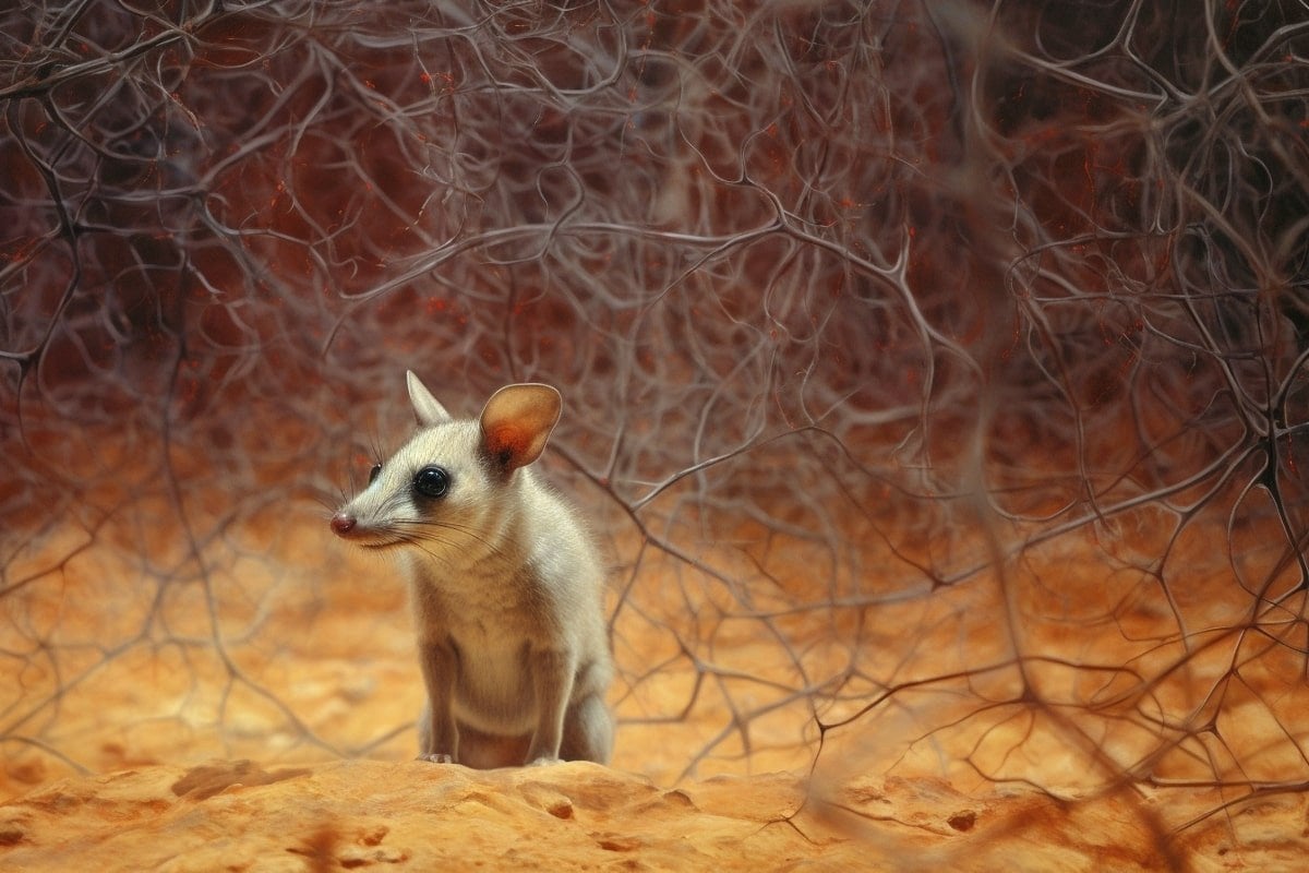
Summary: The early stages of human brain development are mirrored in the brains of marsupials.
Researchers observed that the neural activity patterns in the Australian fat-tailed dunnart, a marsupial species, resemble those in the human brain during gestation.
These findings could help improve our understanding of neurodevelopmental disorders such as autism spectrum disorder (ASD). The marsupial brain development study also provides an unprecedented opportunity to delve into the earliest stages of brain evolution.
Key Facts:
- Marsupial brain development, which mostly occurs postnatally, is similar to human brain development in utero.
- Researchers used light indicators to record the electrical activity of neurons in marsupial joeys, revealing distinct patterns.
- The study suggests that subtle defects in these brain activity patterns could contribute to neurodevelopmental conditions like ASD.
Source: University of Queensland
University of Queensland research has revealed features of early human brain development are mimicked in the brains of marsupials.
Lead author Dr Rodrigo Suárez from UQ’s Queensland Brain Institute and School of Biomedical Sciences, said the finding could lead to a better understanding of brain patterns linked to neurodevelopmental conditions like autism spectrum disorder (ASD).
“Marsupials are mammals born at extremely early stages – the equivalent to mid-gestation in human terms,” Dr Suárez said.
“Most marsupial brain development happens postnatally, inside the mother’s pouch.
“Because of this, we’ve been able to study patterns of neural activity in the Australian native fat-tailed dunnart and found they’re similar to those in the human brain in utero.”

The research used light indicators to record the electrical activity of neurons in marsupial joeys.
“We followed the onset and maturation of complex activity patterns, using advanced microscopy to read how the joey’s developing brain cells first communicate,” Dr Suárez said.
“There were distinct patterns from the outset indicating not only that neural activity begins before sensory experience, but that unique electrical features in newborn cells might be crucial for the healthy establishment of brain connections.
“Likewise, subtle defects in these patterns could lead to neurodevelopmental conditions like ASD.”
Dr Suárez said it was well established that human babies respond to stimulation well before birth.
“But exactly when, where and how electrical activity begins in the developing brain has remained largely unknown,” he said.
“This is mostly because only mammals have evolved a cerebral cortex – the wrinkly surface of our brains that controls sensory motor and cognitive tasks – and most experimental models can’t survive at such early stages outside the uterus.”
Dr Suárez said studying marsupials could help researchers go further back in brain evolution.
“These findings highlight early processes of brain development that arose millions of years ago, and are ongoing with little change, likely influencing the evolution and diversification of the cerebral cortex.”
About this neurodevelopment research news
Author: Merrett Pye
Source: University of Queensland
Contact: Merrett Pye – University of Queensland
Image: The image is credited to Neuroscience News
Original Research: Closed access.
“Cortical activity emerges in region-specific patterns during early brain development” by Rodrigo Suárez et al. PNAS
Abstract
Cortical activity emerges in region-specific patterns during early brain development
The development of precise neural circuits in the brain requires spontaneous patterns of neural activity prior to functional maturation. In the rodent cerebral cortex, patchwork and wave patterns of activity develop in somatosensory and visual regions, respectively, and are present at birth.
However, whether such activity patterns occur in noneutherian mammals, as well as when and how they arise during development, remain open questions relevant for understanding brain formation in health and disease.
Since the onset of patterned cortical activity is challenging to study prenatally in eutherians, here we offer an approach in a minimally invasive manner using marsupial dunnarts, whose cortex forms postnatally.
We discovered similar patchwork and travelling waves in the dunnart somatosensory and visual cortices at stage 27 (equivalent to newborn mice) and examined earlier stages of development to determine the onset of these patterns and how they first emerge.
We observed that these patterns of activity emerge in a region-specific and sequential manner, becoming evident as early as stage 24 in somatosensory and stage 25 in visual cortices (equivalent to embryonic day 16 and 17, respectively, in mice), as cortical layers establish and thalamic axons innervate the cortex.
In addition to sculpting synaptic connections of existing circuits, evolutionarily conserved patterns of neural activity could therefore help regulate other early events in cortical development.


