

Summary: A new language-switching experiment revealed traditional categorization of brain areas may not be sufficient. Researchers set their sights on the caudal inferior parietal cortex to better understand functional categorization in the brain.
Source: Leiden University
Based on the results of a language-switching experiment, Ph.D. candidate Fatemeh (Simeen) Tabassi Mofrad MA and Professor Niels Schiller have discovered that the traditional categorization of brain areas is not sufficient.
They published their research findings in the scientific journal NeuroImage.
“The traditional picture of brain areas was that different parts of the cortex are either task-related or resting state-related,” Tabassi Mofrad explains.
By looking at the connectivity patterns of the caudal inferior parietal cortex (IPC), however, the neurolinguists found a new functional brain category that has modulating functions; this brain area displays no similarities to task-related cortical areas, nor to brain areas that are active when the brain is not processing external stimuli.
More complete picture of the brain
The researchers’ findings lead to a more complete picture of the brain. “The threefold function of the IPC was ignored for a long time in earlier studies, because this brain area was viewed as a single entity when describing functions of the IPC,” says Tabassi Mofrad.
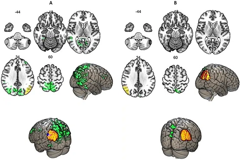
“However, the three sub-sections of this cortical area have different characteristics from each other, and what previous studies reported about how the IPC processes cognitive tasks is not representative of all three of its components. This led to inconsistences in the existing literature about the functions of the IPC.”
Ultimately, Tabassi Mofrad expects her research to have an impact on clinical neuroscience in the near future. She herself will first expand and continue her current research. “I have already mapped the connectivity patterns of the middle IPC and will continue to further investigate the modulating cortical areas.”
About this neuroscience research news
Author: Press Office
Source: Leiden University
Contact: Press Office – Leiden University
Image: The image is credited to NeuroImage
Original Research: Open access.
“Mapping caudal inferior parietal cortex supports the hypothesis about a modulating cortical area” by Fatemeh Tabassi Mofrad et al. NeuroImage
Abstract
Mapping caudal inferior parietal cortex supports the hypothesis about a modulating cortical area
The cytoarchitectonically tripartite organization of the inferior parietal cortex (IPC) into the rostral, the middle and the caudal clusters has been generally ignored when associating different functions to this part of the cortex, resulting in inconsistencies about how IPC is understood. In this study, we investigated the patterns of functional connectivity of the caudal IPC in a task requiring cognitive control, using multiband EPI.
This part of the cortex demonstrated functional connectivity patterns dissimilar to a cognitive control area and at the same time the caudal IPC showed negative functional associations with both task-related brain areas and the precuneus cortex, which is active during resting state.
We found evidence suggesting that the traditional categorization of different brain areas into either task-related or resting state-related networks cannot accommodate the functions of the caudal IPC.
This underlies the hypothesis about a new brain functional category as a modulating cortical area proposing that its involvement in task performance, in a modulating manner, is marked by deactivation in the patterns of functional associations with parts of the brain that are recognized to be involved in doing a task, proportionate to task difficulty; however, its patterns of functional connectivity in some other respects do not correspond to the resting state-related parts of the cortex.


