Cobwebs clear around how body remembers, fights infection.
Scientists at the Virginia Tech Carilion Research Institute have found a potential way to influence long-term memory formation in the immune system. The researchers published their results this week in Nature Communications.
“We’ve found that the inherent flexibility of the immune system is even more complex than previously understood,” said Kenneth Oestreich, senior author of the paper and an assistant professor at the Virginia Tech Carilion Research Institute.
At the first hint of a viral or bacterial invasion, the immune system launches an orchestrated front-line defense.
If infection manages to take hold, the immune system creates specialized cells to combat the specific invading pathogen. These cells, known as effector cells, encounter and begin to destroy the specific pathogen.
Once the infection has been eliminated, some of those cells linger in the body to become memory cells in the immune system. If the host becomes re-infected with the same bug, the immune system will recognize and quickly respond with a targeted attack.
“That’s the basis of vaccine-mediated immunity,” said Paul McDonald, a research scientist at the Virginia Tech Carilion Research Institute and first author on the paper. “But how these memory cells arise after an infection has always been a bit of a mystery. Our study suggests that these important cells may arise directly from the effector population.”
In this study, the researchers learned that proteins responding to the cells’ environment are able to influence the memory gene program. The environment can actually push the effector cells towards memory.
“It was once believed that effector and memory cells arose as two distinct populations, with some cells initially fated to be effector type and some to be memory,” McDonald said. “We’ve now seen that there is much more fluidity between the cell types than originally thought.”
The presence of certain proteins can influence the cell’s fate. Interleukin-2, for instance, is a highly inflammatory protein produced at the start of an infection.
Interleukin-2 levels remain high until the infection is under control. As the interleukin-2 levels drop, effector cells can turn on a protein called Bcl-6. This protein initiates the production of T follicular helper cells. These cells work in the immune system to generate antibodies specifically against the invading pathogen.
“We had previously discovered that in a low interleukin-2 environment, the TFH gene program turns on, apparently pushing effector cells to become T follicular helper cells,” Oestreich said. “In this study, we realized that both the T follicular helper cells and memory profiles were turning on.”
Memory cells can recognize pathogens that have previously infected the body and quickly respond with the correct antibodies.
“We found that the effector cells not only have both the T follicular helper cells and central memory profiles, but they also expressed receptors for two different proteins — interleukin-6 and interleukin-7,” Oestreich said.
Scientists already knew interleukin-6 influenced T follicular helper cell development, and they also knew interleukin-7 was important for memory maintenance, ensuring memory cells persist for years after an initial infection. Oestreich’s team discovered that the presence of either of these proteins could push a cell toward one fate or another.
“Just as the presence of interleukin-2 or interleukin-6 increases Bcl-6 and encourages the T follicular helper cell gene program, the presence of interleukin-7 can decrease Bcl-6 and influence effector cells to become memory cells,” McDonald said.
Memory cells conduct antigen surveillance and are immediately able to recognize the pathogen.

“Central memory cells allow for such a quick response to antigens that infection typically doesn’t have a chance to form. You might not even know you’ve been infected,” McDonald said. “Formation of these cells is critical for a vaccine to work well.”
Vaccines introduce antigens to the immune system to trigger antibody and memory cell production, usually without the host developing symptoms of an illness. If an active antigen invades the body, the memory cells can reactivate and assist in antibody production, eliminating the infection before it causes symptoms.
“We still have a lot of work to do,” Oestreich said. “But this is a promising first step in understanding the mechanisms by which memory cells form, which ultimately will play a role in improving vaccine efficacy.
Source: Ashley WennersHerron – Virginia Tech
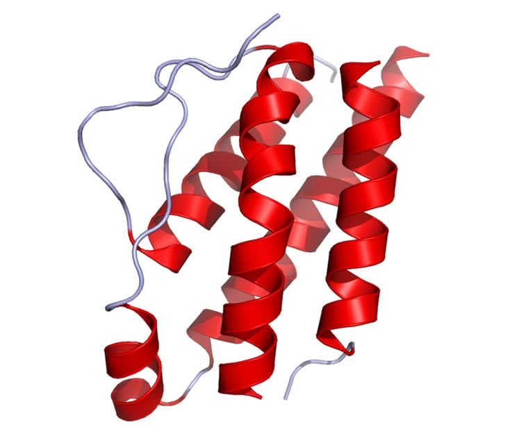
Image Source: The image is credited to Ramin Herati and is in the public domain
Original Research: Full open access research for “IL-7 signalling represses Bcl-6 and the TFH gene program” by Paul W. McDonald, Kaitlin A. Read, Chandra E. Baker, Ashlyn E. Anderson, Michael D. Powell, André Ballesteros-Tato and Kenneth J. Oestreich is in Nature Communications. Published online January 8 2016 doi:10.1038/ncomms10285
Abstract
IL-7 signalling represses Bcl-6 and the TFH gene program
The transcriptional repressor Bcl-6 is linked to the development of both CD4+ T follicular helper (TFH) and central memory T (TCM) cells. Here, we demonstrate that in response to decreased IL-2 signalling, T helper 1 (TH1) cells upregulate Bcl-6 and co-initiate TFH- and TCM-like gene programs, including expression of the cytokine receptors IL-6Rα and IL-7R. Exposure of this potentially bi-potent cell population to IL-6 favours the TFH gene program, whereas IL-7 signalling represses TFH-associated genes including Bcl6 and Cxcr5, but not the TCM-related genes Klf2 and Sell. Mechanistically, IL-7-dependent activation of STAT5 contributes to Bcl-6 repression. Importantly, antigen-specific IL-6Rα+IL-7R+ CD4+ T cells emerge from the effector population at late time points post influenza infection. These data support a novel role for IL-7 in the repression of the TFH gene program and evoke a divergent regulatory mechanism by which post-effector TH1 cells may contribute to long-term cell-mediated and humoral immunity.
“IL-7 signalling represses Bcl-6 and the TFH gene program” by Paul W. McDonald, Kaitlin A. Read, Chandra E. Baker, Ashlyn E. Anderson, Michael D. Powell, André Ballesteros-Tato and Kenneth J. Oestreich is in Nature Communications. Published online January 8 2016 doi:10.1038/ncomms10285


