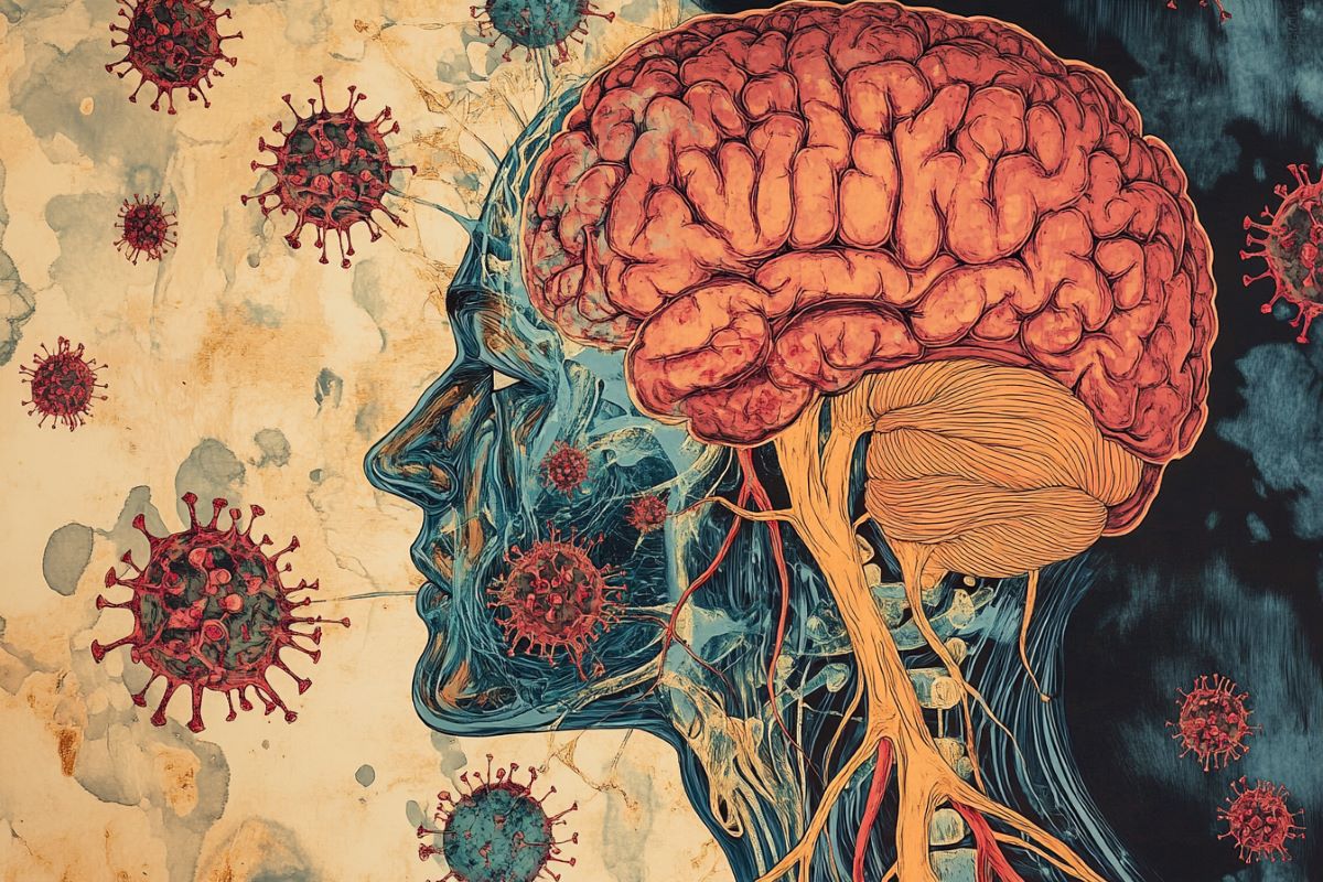
Summary: Researchers have linked chronic intestinal infections caused by cytomegalovirus (HCMV) to a unique subtype of Alzheimer’s disease. The virus may travel from the gut to the brain via the vagus nerve, altering immune responses and contributing to hallmark Alzheimer’s changes like amyloid plaques and tau tangles.
While HCMV infection is common and typically harmless, this study found that it may cause chronic brain inflammation in certain individuals. The researchers demonstrated how HCMV induces molecular changes associated with Alzheimer’s in human brain cell models.
These findings highlight the potential for antiviral therapies to address this subtype of Alzheimer’s. Ongoing work aims to develop a blood test to identify individuals with chronic HCMV infection and evaluate treatment options.
Key Facts:
- Gut-to-Brain Link: HCMV infections in the gut may reach the brain via the vagus nerve, contributing to Alzheimer’s.
- Molecular Impact: The virus triggers amyloid and tau production, leading to neuron damage.
- Therapeutic Potential: Researchers are exploring antiviral drugs to treat this Alzheimer’s subtype.
Source: Arizona State University
Arizona State University and Banner Alzheimer’s Institute researchers, along with their collaborators, have discovered a surprising link between a chronic gut infection caused by a common virus and the development of Alzheimer’s disease in a subset of people.
It is believed most humans are exposed to this virus — called cytomegalovirus or HCMV — during the first few decades of life. Cytomegalovirus is one of nine herpes viruses, but it is not considered a sexually transmitted disease. The virus is usually passed through exposure to bodily fluids and spread only when the virus is active.
Video Credit: Neuroscience News
According to the new research, in some people, the virus may linger in an active state in the gut, where it may travel to the brain via the vagus nerve — a critical information highway that connects the gut and brain. Once there, the virus can change the immune system and contribute to other changes associated with Alzheimer’s disease.
If the researchers’ hypotheses are confirmed, they may be able to evaluate whether existing antiviral drugs could treat or prevent this form of Alzheimer’s disease. They are currently developing a blood test to identify people who have an active HCMV infection and who might benefit from antiviral medication.
“We think we found a biologically unique subtype of Alzheimer’s that may affect 25% to 45% of people with this disease,” said Dr. Ben Readhead, co-first author of the study and research associate professor with ASU-Banner Neurodegenerative Disease Research Center in the Biodesign Institute at ASU.
“This subtype of Alzheimer’s includes the hallmark amyloid plaques and tau tangles—microscopic brain abnormalities used for diagnosis—and features a distinct biological profile of virus, antibodies and immune cells in the brain.”
The findings were published today in “Alzheimer’s & Dementia: The Journal of the Alzheimer’s Association.”
Researchers from ASU, Banner Alzheimer’s Institute, Banner Sun Health Research Institute, and the Translational Genomics Research Institute (TGen) led the collaborative effort, which included investigators with UMass Chan Medical School, Institute for Systems Biology, Rush University Medical Center, Icahn School of Medicine at Mount Sinai, and other institutions.
The research team suggests that some people exposed to HCMV develop a chronic intestinal infection. The virus then enters the bloodstream or travels through the vagus nerve to the brain.
There, it is recognized by the brain’s immune cells, called microglia, which turn on the expression of a specific gene called CD83. The virus may contribute to the biological changes involved in the development of Alzheimer’s.
The role of the brain’s immune cells
Microglia, or the brain’s immune cells, are activated when responding to infections. While initially protective, a sustained increase in microglial activity may lead to chronic inflammation and neuronal damage, which is implicated in the progression of neurodegenerative diseases, including Alzheimer’s.
In a study published earlier this year in “Nature Communications,” the researchers found that the postmortem brains of research participants with Alzheimer’s disease were more likely than those without Alzheimer’s to harbor specifically CD83(+) microglia.
While exploring why this occurred, they discovered an antibody in the intestines of these subjects — consistent with the possibility that an infection could contribute to this form of Alzheimer’s.
In the newest study, investigators sought to understand what might be driving the intestinal antibody production. The team examined spinal fluid from these same individuals, which revealed that the antibodies were specifically against HCMV. This prompted a search for evidence of HCMV infection in the intestine and brain tissue of these subjects – which they found.
They also saw HCMV within the vagus nerve of the same subjects, raising the possibility that this is how the virus travels to the brain. Working with RUSH University, the researchers were able to reproduce the association between cytomegalovirus infection and CD83(+) microglia in an independent cohort of Alzheimer’s patients.
To further investigate the impact of this virus, the research team then used human brain cell models to demonstrate the virus’s ability to induce molecular changes related to this specific form of Alzheimer’s disease. Exposure to the virus did increase the production of amyloid and phosphorylated tau proteins and contributed to the degeneration and death of neurons.
Is HCMV to blame for Alzheimer’s disease in some people?
HCMV can infect humans of all ages. In most healthy individuals, infection occurs without symptoms but may present as a mild, flu-like illness. About 80% of people show evidence of antibodies by age 80.
Nonetheless, the researchers detected intestinal HCMV only in a subset of individuals, and this infection seems to be a relevant factor in the presence of the virus in the brain. For this reason, the researchers note that simply coming into contact with HCMV, which happens to almost everyone, should not be cause for concern.
And, although researchers proposed more than 100 years ago that harmful viruses or microbes could contribute to Alzheimer’s disease, no single pathogen has consistently been linked to the disease.
The researchers propose these two studies illustrate the potential impact that infections can have on brain health and neurodegeneration broadly. Yet, they add that independent studies are needed to put their findings and resulting hypotheses to the test.
The NOMIS Foundation, Banner Alzheimer’s Foundation, National Institutes of Health, and Arizona Alzheimer’s Consortium supported the study. Arizona’s unique biorepositories, particularly the Brain and Body Donation Program at Banner Sun Health Research Institute, provided tissue samples and resources, including the colon, vagus nerve, brain and spinal fluid.
Rush University-led Religious Orders Study and Memory and Aging Study provided additional brain samples and data. This allowed researchers to conduct a more nuanced investigation, highlighting the systemic rather than purely neurological roots of Alzheimer’s disease.
“It was critically important for us to have access to different tissues from the same individuals. That allowed us to piece the research together. Arizona is the only place I know of where a study like this could have been done, and we’re grateful to the Banner Health Brain and Body Donation Program for its support,” said Readhead, also the Edson Endowed Professor of Dementia Research at the center.
“We are extremely grateful to our research participants, colleagues, and supporters for the chance to advance this research in a way that none of us could have done on our own,” said Dr. Eric Reiman, Executive Director of Banner Alzheimer’s Institute and the study’s senior author.
“We’re excited about the chance to have researchers test our findings in ways that make a difference in the study, subtyping, treatment and prevention of Alzheimer’s disease.”
The findings of the recent study raise an important question: Could antiviral medications help treat Alzheimer’s patients who have a chronic HCMV infection?
The investigators are working now on a blood test to identify individuals with this type of chronic intestinal HCMV infection. They hope to use it in conjunction with emerging Alzheimer’s blood tests to evaluate whether existing antiviral drugs could be used to treat or prevent this form of Alzheimer’s disease.
Research institutions involved in the study, published in the journal Alzheimer’s & Dementia: ASU-Banner Neurodegenerative Disease Research Center in the Biodesign Institute at ASU; Weill Cornell Medicine; Icahn School of Medicine; University of Massachusetts Chan Medical School; The Translational Genomics Research Institute; Institute for Systems Biology; Serimmune, Inc; Rush University Medical Center; Banner Sun Health Research Institute; and Banner Alzheimer’s Institute.
About this microbiome and Alzheimer’s disease research news
Author: Sandy Keaton Leander
Source: Arizona State University
Contact: Sandy Keaton Leander – Arizona State University
Image: The image is credited to Neuroscience News
Original Research: Open access.
“Alzheimer’s disease-associated CD83(+)microglia are linked with increased immunoglobulin G4 and human cytomegalovirus in the gut, vagal nerve, and brain” by Ben Readhead et al. Alzheimer’s & Dementia
Abstract
Alzheimer’s disease-associated CD83(+)microglia are linked with increased immunoglobulin G4 and human cytomegalovirus in the gut, vagal nerve, and brain
INTRODUCTION
While there may be microbial contributions to Alzheimer’s disease (AD), findings have been inconclusive. We recently reported an AD-associated CD83(+) microglia subtype associated with increased immunoglobulin G4 (IgG4) in the transverse colon (TC).
METHODS
We used immunohistochemistry (IHC), IgG4 repertoire profiling, and brain organoid experiments to explore this association.
RESULTS
CD83(+) microglia in the superior frontal gyrus (SFG) are associated with elevated IgG4 and human cytomegalovirus (HCMV) in the TC, anti-HCMV IgG4 in cerebrospinal fluid, and both HCMV and IgG4 in the SFG and vagal nerve. This association was replicated in an independent AD cohort. HCMV-infected cerebral organoids showed accelerated AD pathophysiological features (Aβ42 and pTau-212) and neuronal death.
DISCUSSION
Findings indicate complex, cross-tissue interactions between HCMV and the adaptive immune response associated with CD83(+) microglia in persons with AD. This may indicate an opportunity for antiviral therapy in persons with AD and biomarker evidence of HCMV, IgG4, or CD83(+) microglia.


