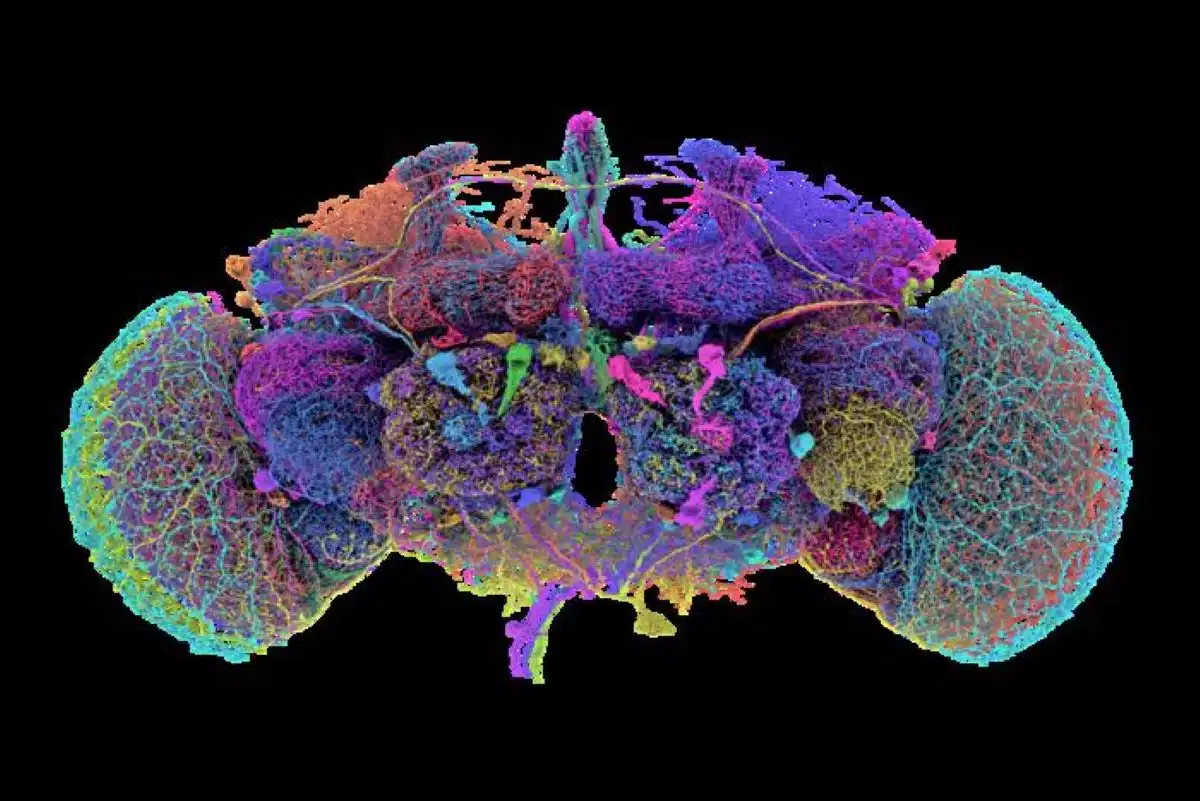
Summary: Researchers have created the first neuron-by-neuron map of an adult fruit fly brain, marking a major achievement in brain mapping. The map, built using 21 million images, connects nearly 140,000 neurons and 50 million synapses, making it the most complex brain map of any adult animal to date.
This “connectome” provides a roadmap for understanding neural circuits and behaviors in more complex species, including humans. The development, powered by AI, opens doors for new insights into brain function and diseases.
Key Facts:
- The adult fruit fly brain contains 140,000 neurons and 50 million synapses.
- The map was built from 21 million images using AI to process neural data.
- Fruit flies share 60% of human DNA, aiding research on human brain diseases.
Source: Princeton
A Princeton-led team of scientists has built the first neuron-by-neuron and synapse-by-synapse roadmap through the brain of an adult fruit fly (Drosophila melanogaster), marking a major milestone in the study of brains.
This research is the flagship article in the Oct. 2 special issue of Nature, which is devoted to the new fruit fly “connectome.”
Previous researchers mapped the brain of a C. elegans worm, with its 302 neurons, and the brain of a larval fruit fly, which had 3,000 neurons, but the adult fruit fly is several orders of magnitude more complex, with almost 140,000 neurons and roughly 50 million synapses connecting them.
Fruit flies share 60% of human DNA, and three in four human genetic diseases have a parallel in fruit flies. Understanding the brains of fruit flies is a steppingstone to understanding brains of larger more complex species, like humans.
“This is a major achievement,” said Mala Murthy, director of the Princeton Neuroscience Institute and, with Sebastian Seung, co-leader of the research team. “There is no other full brain connectome for an adult animal of this complexity.” Murthy is also Princeton’s Karol and Marnie Marcin ‘96 Professor of Neuroscience.
Princeton’s Seung and Murthy are co-senior authors on the flagship paper of the Nature issue, which includes a suite of nine related papers with overlapping sets of authors, led by researchers from Princeton University, the University of Vermont, the University of Cambridge, the University of California-Berkeley, UC-Santa Barbara, Freie Universität-Berlin, and the Max Planck Florida Institute for Neuroscience.
The work was funded in part by the NIH’s BRAIN Initiative, the Princeton Neuroscience Institute’s Bezos Center for Neural Circuit Dynamics and McDonnell Center for Systems Neuroscience, and other public and private neuroscience institutes and funds, listed at the end of this document.
The map was developed by the FlyWire Consortium, which is based at Princeton University and made up of teams in more than 76 laboratories with 287 researchers around the world as well as volunteer gamers.
Sven Dorkenwald, the lead author on the flagship Nature paper, spearheaded the FlyWire Consortium.
“What we built is, in many ways, an atlas,” said Dorkenwald, a 2023 Ph.D. graduate of Princeton now at the University of Washington and the Allen Institute for Brain Science.
“Just like you wouldn’t want to drive to a new place without Google Maps, you don’t want to explore the brain without a map. What we have done is build an atlas of the brain, and added annotations for all the businesses, the buildings, the street names. With this, researchers are now equipped to thoughtfully navigate the brain as we try to understand it.”
And just like a map that traces out every tiny alley as well as every superhighway, the fly connectome shows connections within the fruit fly brain at every scale.
The map was built from 21 million images taken of a female fruit fly brain by a team of scientists led by Davi Bock, then at the Howard Hughes Medical Institute’s Janelia Research Campus and now at the University of Vermont.
Using an AI model built by researchers and software engineers working with Princeton’s Sebastian Seung, the lumps and blobs in those images were turned into a labeled, three-dimensional map. Instead of keeping their data confidential, the researchers opened their in-progress neural map to the scientific community from the beginning.
“Mapping the whole brain has been made possible by advances in AI computing. It would have not been possible to reconstruct the entire wiring diagram manually. This is a display of how AI can move neuroscience forward,’ said Prof. Sebastian Seung, one of the co-leaders of the research and Princeton’s Evnin Professor in Neuroscience and a professor of computer science.
“Now that we have this brain map, we can close the loop on which neurons relate to which behaviors,” said Dorkenwald.
The development could lead to tailored treatments to brain diseases.
“In many respects, it (the brain) is more powerful than any human-made computer, yet for the most part we still do not understand its underlying logic,” said John Ngai, director of the U.S. National Institutes of Health’s BRAIN Initiative, which provided partial funding for the FlyWire project.
“Without a detailed understanding of how neurons connect with one another, we won’t have a basic understanding of what goes right in a healthy brain or what goes wrong in disease.”
Funding: This research was supported by the National Institutes of Health (NIH) BRAIN Initiative (RF1 MH117815, RF1 MH129268, 1RF1MH120679-01 and U24 NS126935) and National Institute Of Neurological Disorders And Stroke (NINDS) (RF1NS121911); the Princeton Neuroscience Institute’s Bezos Center for Neural Circuit Dynamics and McDonnell Center for Systems Neuroscience; Google; the Allen Institute for Brain Science; the National Science Foundation (NSF Neuronex2 2014862, Neuronex2 MRC MC_EX_MR/T046279/1, MRC MC-U105188491, PHY-1734030); Wellcome Trust Collaborative Award (203261/Z/16/Z and 220343/Z/20/Z); a Marie Skłodowska-Curie postdoctoral fellowship (H2020-WF-01-2018-867459); the Portuguese Research Council (Grant PTDC/MED-NEU/4001/2021); and the Intelligence Advanced Research Projects Activity (IARPA) via the Department of Interior (DOI) (D16PC0005).
About this brain mapping research news
Author: Liz Fuller-Wright
Source: Princeton
Contact: Liz Fuller-Wright – Princeton
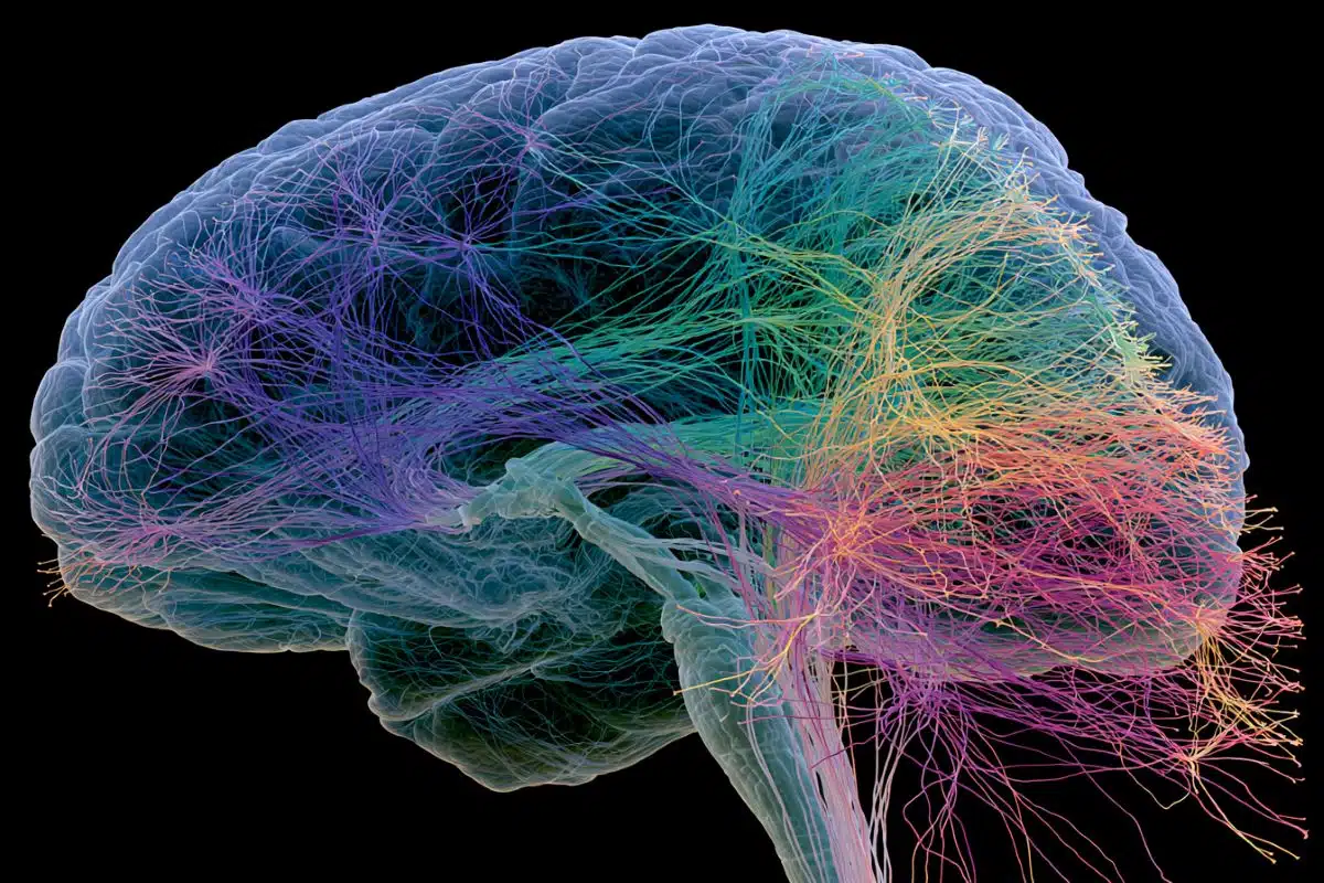
Image: The image is credited to Tyler Sloan and Amy Sterling / FlyWire / Princeton University
Original Research: Open access.
“Neuronal wiring diagram of an adult brain” by Mala Murthy et al. Nature
Abstract
Neuronal wiring diagram of an adult brain
Connections between neurons can be mapped by acquiring and analysing electron microscopic brain images. In recent years, this approach has been applied to chunks of brains to reconstruct local connectivity maps that are highly informative, but nevertheless inadequate for understanding brain function more globally.
Here we present a neuronal wiring diagram of a whole brain containing 5 × 10⁷ chemical synapses between 139,255 neurons reconstructed from an adult female Drosophila melanogaster. The resource also incorporates annotations of cell classes and types, nerves, hemilineages and predictions of neurotransmitter identities.
Data products are available for download, programmatic access and interactive browsing and have been made interoperable with other fly data resources.
We derive a projectome—a map of projections between regions—from the connectome and report on tracing of synaptic pathways and the analysis of information flow from inputs (sensory and ascending neurons) to outputs (motor, endocrine and descending neurons) across both hemispheres and between the central brain and the optic lobes.
Tracing from a subset of photoreceptors to descending motor pathways illustrates how structure can uncover putative circuit mechanisms underlying sensorimotor behaviours.
The technologies and open ecosystem reported here set the stage for future large-scale connectome projects in other species.