

Summary: Researchers unveiled the most comprehensive connectome of the adult fruit fly nerve cord, analogous to the human spinal cord, providing an exceptional resource for the scientific community.
The connectome, constructed from about 23,000 neurons, reveals the intricate network controlling the fly’s motor functions. New insights have already emerged from the data, challenging previous theories on fly movement.
This achievement not only advances understanding of fruit fly neurology, but also serves as a model for similar future projects.
Key Facts:
- The fruit fly nerve cord connectome includes about 23,000 neurons, 10 million pre-synaptic sites, and 74 million post-synaptic densities.
- It’s the most in-depth and complete connectome of an adult fruit fly nerve cord to date.
- The connectome has already revealed that some behaviors involving the same muscles use distinct pre-motor microcircuits, contradicting existing theories.
Source: Janelia Research Campus
Already this year, researchers have released a wiring diagram of the larval fruit fly brain. Connectomes of the complete adult female fly brain and the optic lobe are expected in 2023, with the complete male fly nervous system connectome following soon.
On June 6, Janelia scientists and collaborators in the US and UK added another piece to the connectome puzzle with the unveiling of the wiring diagram of the male adult nerve cord, dubbed the MANC.
The connectome, a joint effort by Janelia’s FlyEM Project Team and collaborators, is detailed in preprints on bioRxiv and is freely available to researchers worldwide through Janelia websites.
With about 23,000 neurons, 10 million pre-synaptic sites, and 74 million post-synaptic densities, the MANC is the most in-depth and complete connectome of an adult fruit fly nerve cord – a structure analogous to the human spinal cord that controls most of the fly’s motor functions.
The unprecedented detail in this map of neurons and their connections will help scientists figure out how a fly moves its legs or flaps its wings.
If the 23,000 neurons making up the MANC connectome were laid end-to-end, they would stretch for about 44 meters.
Preprints released alongside the connectome data describe the different cell types, their origins and connections, and the biological insights starting to emerge from the data. The fruit fly is a key organism that neuroscientists use to probe how the nervous system works, so having connectomes is critical for uncovering how cells work together to enable behavior.
“Once you can see a whole network, you can start to ask big organizational questions,” says Gwyneth Card, an HHMI Investigator at Columbia University’s Zuckerman Institute and former Janelia group leader, who helped lead the project.
The MANC and the other connectomes being released follow in the footsteps of the hemibrain connectome released by Janelia scientists in 2020. At the time, the hemibrain – a portion of the adult fly brain – was the largest and most comprehensive wiring diagram ever completed, showing that a feat many thought impossible could be done.
The release of the hemibrain led to additional support for and interest in connectome efforts. Researchers are now filling in pieces missing from the hemibrain, and the goal of mapping the entire central nervous system of both a male and female adult fruit fly is within reach.
“This train is going to keep rolling,” Card says. “You are just seeing the beginning.”
Constructing the MANC
The MANC connectome was constructed using methods akin to those used to map the hemibrain, with the Janelia team preparing the nerve cord sample and imaging layer after layer of nanometer-thick slices on focused ion beam scanning electron microscopes. Google’s algorithms and computers stitched the images together and did a first pass at identifying neurons.

Then, a team of Janelians and collaborators set about proofreading the data – a manual effort to ensure that the shape and connectivity of neurons are correct, and one of the most time-consuming parts of the process. Because of the COVID-19 pandemic, the team developed software to work on home computers. That, along with additional funding from the Wellcome Trust, meant international collaborators could more easily help with the effort.
“Since it has been completely proofread and we can find all the same neurons on the fly’s left and right, we can tell colleagues, ‘You can trust this,’” says Greg Jefferis, a neuroscientist at the MRC Laboratory of Molecular Biology and University of Cambridge and another project leader who is part of the FlyEM Project Team Steering Committee.
Researchers at Cambridge also identified the different cell types, where they are found along the fly’s body, and from which stem cells they originated, helping to tease out some of the organizational principles.
“The ventral nerve cord has basically been seen as a black box,” says Lisa Marin, a research associate at the University of Cambridge who led the cell-typing effort.
“A large majority of the neurons have never been identified. So a big part of our process was to divide these up into smaller populations and then dig into the connectivity.”
Examination of the connectome data has already started to uncover some surprises. Card and her team found that some behaviors involving the same muscles use distinct pre-motor microcircuits, not the same circuits, as previously thought.
Jefferis and his team described the complex repeated circuits that control the legs and found, surprisingly, that the interconnections coordinating the legs differ from existing models.
Many more insights from the MANC will happen as other researchers start to probe the data, which can be accessed through neuPrint and Clio, online tools developed at Janelia.
“It’s clear that these connectomes are so rich and that they’re really only the starting point for trying to understand how this system works,” Card says. “It will take the whole community to dig in to get the breadth of different behaviors that people study in different contexts, to probe this network. That’s how we are going to tease out the higher principles.”
Along with the scientific insights to be gained, the project also serves as one model for other groups undertaking connectome efforts.
“This kind of cooperation is going to be absolutely necessary when people start moving to the mouse connectome and things like that,” says Lou Scheffer, a principal scientist at Janelia and a member of the FlyEM team.
“There’s no conceivable way any single organization could do it, and so this is a prototype for that sort of cooperation.”
Datasets: https://www.janelia.org/project-team/flyem/manc-connectome
About this neuroscience research news
Author: Gwyneth Card
Source: Janelia Research Campus
Contact: Gwyneth Card – Janelia Research Campus
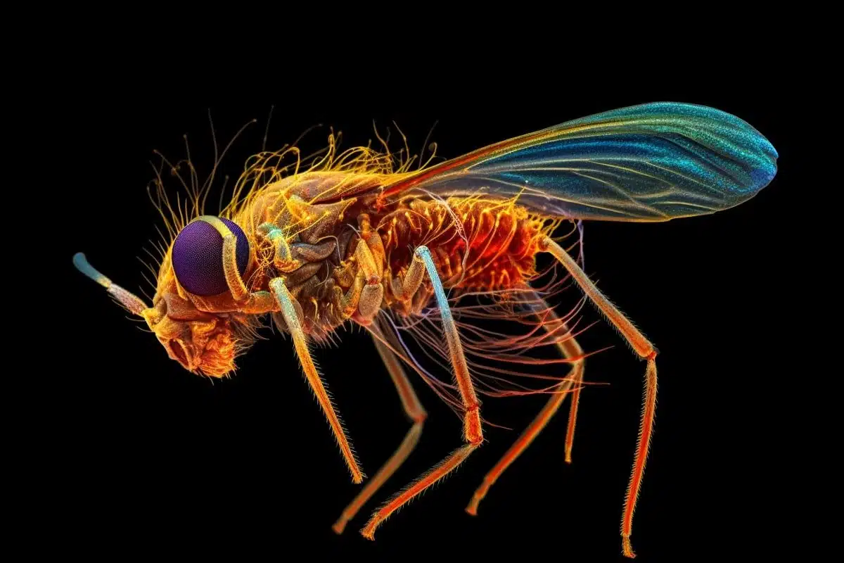
Image: The top image is credited to Neuroscience News. The article image is credited to FlyEM/Janelia Research Campus
Original Research: Closed access.
“A Connectome of the Male Drosophila Ventral Nerve Cord” by Shin-ya Takemura et al. bioRXiv
Closed access.
“Systematic annotation of a complete adult male Drosophila nerve cord connectome reveals principles of functional organisation” by Elizabeth C. Marin et al. bioRXiv
Abstract
A Connectome of the Male Drosophila Ventral Nerve Cord
Animal behavior is principally expressed through neural control of muscles. Therefore understanding how the brain controls behavior requires mapping neuronal circuits all the way to motor neurons.
We have previously established technology to collect large-volume electron microscopy data sets of neural tissue and fully reconstruct the morphology of the neurons and their chemical synaptic connections throughout the volume. Using these tools we generated a dense wiring diagram, or connectome, for a large portion of the Drosophila central brain.
However, in most animals, including the fly, the majority of motor neurons are located outside the brain in a neural center closer to the body, i.e. the mammalian spinal cord or insect ventral nerve cord (VNC).
In this paper, we extend our effort to map full neural circuits for behavior by generating a connectome of the VNC of a male fly.
Abstract
Systematic annotation of a complete adult male Drosophila nerve cord connectome reveals principles of functional organisation
Our companion paper (Takemura et al., 2023) introduces the first completely proofread connectome of the nerve cord of an animal that can walk or fly. The base connectome consists of neuronal morphologies and the connections between them.
However, in order to efficiently navigate and understand this connectome, it is crucial to have a system of annotations that systematically categorises and names neurons, linking them to the existing literature.
In this paper we describe the comprehensive annotation of the VNC connectome, first by a system of hierarchical coarse annotations, then by grouping left-right and serially homologous neurons and eventually by defining systematic cell types for the intrinsic interneurons and sensory neurons of the VNC; descending and motor neurons are typed in (Cheong et al., 2023).
We assign a sensory modality to over 5000 sensory neurons, cluster them by connectivity, and identify serially homologous cell types and a layered organisation likely corresponding to peripheral topography. We identify the developmental neuroblast of origin of the large majority of VNC neurons and confirm that (in most cases) all secondary neurons of each hemilineage express a single neurotransmitter.
Neuroblast hemilineages are serially repeated along the segments of the nerve cord and generally exhibit consistent hemilineage-to-hemilineage connectivity across neuromeres, supporting the idea that hemilineages are a major organisational feature of the VNC.
We also find that more than a third of individual neurons belong to serially homologous cell types, which were crucial for identifying motor neurons and sensory neurons across leg neuropils. Categorising interneurons by their neuropil innervation patterns provides an additional organisation axis.
Over half of the intrinsic neurons of the VNC appear dedicated to the legs, with the majority restricted to single leg neuropils; in contrast, inhibitory interneurons connecting different leg neuropils, especially those crossing the midline, appear rarer than anticipated by standard models of locomotor circuitry.
Our annotations are being released as part of the neuprint.janelia.org web application and also serve as the basis of programmatic analysis of the connectome through dedicated tools that we describe in this paper.


