Summary: Some patients with a disorder called hemi-PMO see distortions in the same half of a person’s face, regardless of the angle they view the face. Findings suggest the visual system standardizes all the faces we perceive using the same process, so they can be better compared to faces we have seen before.
Source: Dartmouth College
Imagine if every time you looked at a face, one side of the face always appeared distorted as if it were melting, resembling a painting by Salvador Dalí. This is the case for people who have a rare condition known as hemi-prosopometamophosia (hemi-PMO), which makes looking at faces discomforting. According to a new study published in Current Biology, some people with hemi-PMO see distortions to the same half of a person’s face regardless of how the face is viewed. The results demonstrate that our visual system standardizes all the faces we perceive using the same process so they can be better compared to faces we have seen before.
“Every time we see a face, the brain adjusts our representation of that face so its size, viewpoint, and orientation is matched to faces stored in memory, just like computer face recognition systems such as those used by Facebook and Google,” explains co-author Brad Duchaine, a professor of psychological and brain sciences and the principal investigator of the Social Perception Lab at Dartmouth College. “By aligning the perceived face with faces stored in memory, it’s much easier for us to determine whether the face is one we’ve seen before,” he added.
Hemi-PMO is a rare disorder that may occur after brain damage. When a person with this condition looks at a face, facial features on one side of the face appear distorted. The existence of hemi-PMO suggests the two halves of the face are processed separately. The condition usually dissipates over time, which makes it difficult to study. As a result, little is known about the condition or what it reveals about how human face processing normally works.
The current study focused on a right-handed man in his early sixties (“Patient A.D.”) with hemi-PMO whose symptoms have persisted for years. Like many with this condition, his distortions were caused by damage to a fiber bundle called the splenium that connects visual areas in the left hemisphere and right hemisphere of his brain. Five years ago while A.D. was watching television, he noticed that the right halves of people’s faces looked like they had melted. Yet, the left sides of their faces looked normal. He looked in the mirror at his own face and noticed that the right side of his reflection was also distorted. In contrast, A.D. sees no distortions in other body parts or objects.
The study involved two experiments. In the first, A.D. was presented with images of human faces and non-face images such as objects, houses and cars, and asked to report on distortions. For 17 of the 20 faces, he saw distortions. The distortions were always on the right side of the face and facial features usually appeared to drooped. For example, in one of the faces, A.D. reported that the right eye looked a lot bigger than the left eye while the right eyebrow, right side of the nose, and right side of the lips all hung down unnaturally. Two of the face photographs that did not elicit a distortion showed right profile views in which the right side of the face was not visible. Consistent with his daily experiences, A.D. did not see distortions in any of the non-face images. These results show that his condition affects brain processes specialized for faces.
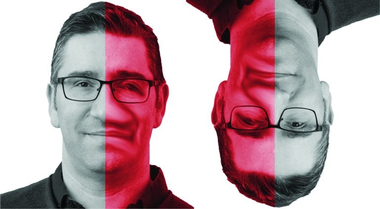
For the second part of the study, A.D. reported on distortions that he saw in 15 different faces that were presented in a variety of ways: in the left and right visual field, at different in-depth rotations, and at four picture plane rotations— 0 degrees or upright, 90 degrees, 180 degrees or upside down, and 270 degrees. Regardless of how the faces were presented, A.D. continued to report that the distortions affected the same facial features. For example, even when a face was presented upside down, A.D. still saw the facial features distorted on the right side of the face even though the distortion now appeared on the left-hand side of the stimulus (the red-shaded region in the faces in the reproduction of Figure 4E below). The consistency of the location of A.D.’s distortion demonstrates that faces, regardless of viewpoint or orientation, are aligned to the same template similar to what computer face recognition systems do. In A.D.’s case, the output from that process is disrupted as it is passed from one brain hemisphere to the other due to his splenium lesion.
About this facial processing research article
Source:
Dartmouth College
Contacts:
Press Office – Dartmouth College
Image Source:
The image is credited to the researchers.
Original Research: Open access
“Face-Specific Perceptual Distortions Reveal A Viewand Orientation-Independent Face Template” by Brad Duchaine et al. Current Biology.
Abstract
Face-Specific Perceptual Distortions Reveal A Viewand Orientation-Independent Face Template
The spatial coordinate system in which a stimulus representation is embedded is known as its reference frame. Every visual representation has a reference frame, and the visual system uses a variety of reference frames to efficiently code visual information [e.g., 1–5]. The representation of faces in early stages of visual processing depends on retino-centered reference frames, but little is known about the reference frames that code the high-level representations used to make judgements about faces. Here, we focus on a rare and striking disorder of face perception—hemi-prosopometamorphopsia (hemi-PMO)—to investigate these reference frames. After a left splenium lesion, Patient A.D. perceives features on the right side of faces as if they had melted. The same features were distorted when faces were presented in either visual field, at different in-depth rotations, and at different picture-plane orientations including upsidedown. A.D.’s results indicate faces are aligned to a view- and orientation-independent face template encoded in a face-centered reference frame, that these face-centered representations are present in both the left and right hemisphere, and that the representations of the left and right halves of a face are dissociable.