

Summary: According to researchers, a hand held device that records electrical activity from the retina may be a useful tool in diagnosis schizophrenia.
Source: Rutgers University.
A portable device common in optometrists’ offices may hold the key to faster diagnosis of schizophrenia, predicting relapse and symptom severity and assessing treatment effectiveness, a Rutgers University study finds.
In the study, published in the May 2018 issue of the Journal of Abnormal Psychology, researchers used RETeval, a hand-held device developed to record electrical activity from the retina, to replicate and extend prior studies showing that people with schizophrenia had abnormal electrical activity in the retina. This was the first time a portable device was used for these tests. The results show the device accurately indicated reduced electrical activity in the retina in multiple cell layers in the participants who had schizophrenia, including in cell types that had not been studied before in this disorder.
“Schizophrenia is a devastating disorder, probably the most disabling disorder long term. Although we know quite a bit about it, it’s still not that well understood,” said Steven Silverstein, professor of psychiatry at Rutgers Robert Wood Johnson Medical School and director of research at Rutgers University Behavioral Health Care (UBHC), who designed the study. “Our study should help generate further research into developing a test that clinicians – like psychologists, psychiatrists or nurses – can use in their offices to diagnose, treat and monitor the condition of people with schizophrenia.”
Looking at biomarkers in the eye as a way to understand psychiatric disorders is a new field of study.
“Since the retina is part of the nervous system, what is happening in the retina is likely reflective of what is occurring in the brain,” Silverstein said. “For example, we know that certain changes in the retina, like thinning tissue [due to cell loss] or weakening electrical activity, occur alongside loss of brain tissue and reduced brain activity in patients with neurological disorders like multiple sclerosis and Parkinson’s disease. We and other researchers are now investigating whether retinal changes are related to brain structure and function changes in schizophrenia.”
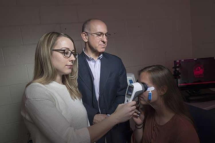
In the just-published study, the researchers evaluated 50 participants: 25 with schizophrenia and 25 with no diagnosed psychiatric disorder. In the test, the participants closed one eye and placed the other against the RETeval device, which flashed 10 to 20 white or colored lights of various intensity against a white or colored background. A tiny skin electrode was placed on the skin under the eye to record the retina’s electrical activity. The participants were tested in normal light and after sitting in the dark for 10 minutes to assess activity in different types of retinal cells. Most individual tests were completed within two minutes.
“Since many of our participants were experiencing severe psychiatric symptoms, such as hallucinations and delusions, we wanted to use a test that was as noninvasive and quick as possible,” Silverstein said.
“While the portable device clearly distinguished people with schizophrenia from those without a psychiatric diagnosis, it’s too soon to call this a diagnostic tool,” said lead author Docia Demmin, a graduate assistant in UBHC’s Division of Schizophrenia Research and a doctoral student in Rutgers Department of Psychology. “However, since every prior study has found that people with schizophrenia exhibit reduced retinal wave forms and slowed retinal responses, our research shows that we closing in on an accurate test that is faster, less invasive, inexpensive and more accessible to patients.”
Source: Patti Verbanas – Rutgers University
Publisher: Organized by NeuroscienceNews.com.
Image Source: NeuroscienceNews.com image is credited to Nick Romanenko / Rutgers University.
Original Research: Abstract for “Electroretinographic anomalies in schizophrenia” by Demmin, Docia L.; Davis, Quentin; Roché, Matthew; and Silverstein, Steven M. in Journal of Abnormal Psychology. Published May 2018.
doi:10.1037/abn0000347
[cbtabs][cbtab title=”MLA”]Rutgers University “Eye Function May Be Key to Schizophrenia Diagnosis.” NeuroscienceNews. NeuroscienceNews, 30 May 2018.
<https://neurosciencenews.com/eye-function-schizophrenia-9180/>.[/cbtab][cbtab title=”APA”]Rutgers University (2018, May 30). Eye Function May Be Key to Schizophrenia Diagnosis. NeuroscienceNews. Retrieved May 30, 2018 from https://neurosciencenews.com/eye-function-schizophrenia-9180/[/cbtab][cbtab title=”Chicago”]Rutgers University “Eye Function May Be Key to Schizophrenia Diagnosis.” https://neurosciencenews.com/eye-function-schizophrenia-9180/ (accessed May 30, 2018).[/cbtab][/cbtabs]
Abstract
Electroretinographic anomalies in schizophrenia
Flash electroretinography (fERG) has been used to identify anomalies in retinal cell function in schizophrenia. Several consistent findings have now emerged, but several potentially important parameters have not yet been investigated. In this study, we recorded light- (photopic) and dark-adapted (scotopic) fERG data from 25 schizophrenia patients and 25 healthy control subjects to (1) determine if past key findings on abnormal photoreceptor and bipolar cell signaling could be replicated; (2) for the first time, examine retinal ganglion cell functioning using the photopic negative response of the fERG; (3) also for the first time, determine responsiveness of schizophrenia patients to a flickering stimulus, as an additional method to isolate cone photoreceptor function; and (4) determine if schizophrenia-related changes in the fERG could be detected using a portable hand-held ERG device. In both photopic and scotopic conditions, schizophrenia patients demonstrated weakened photoreceptor and bipolar cell activations that were most pronounced in response to the most intense stimuli. A reduced cone response to a flicker stimulus and attenuation in ganglion cell activity were also observed in the schizophrenia group. In general, groups did not differ in implicit time of retinal cell responses. These findings (1) replicate and extend prior studies demonstrating reduced photoreceptor (both rod and cone) and bipolar cell functioning in schizophrenia; (2) indicate that retinal ganglion function abnormality can also be detected using fERG; and (3) indicate that these anomalies can be detected using a portable testing device, thereby opening up possibilities for more routine administration of ERG testing.


