

Researchers at The Neuro compile new tool designed to be shared with experts world-wide to accelerate research.
A new detailed map of the hippocampal region of the brain, compiled by researchers at the Montreal Neurological Institute and Hospital-The Neuro at McGill University, is helping the scientific community accelerate research and develop better treatments for patients suffering from epilepsy and other neurological and psychiatric disorders.
The team of researchers, led by Dr. Neda Bernasconi, a neuroscientist specializing in the neuroimaging of epilepsy and co-founder of the Neuroimaging of Epilepsy Laboratory (NOEL) at The Neuro, set out to build and share a detailed model of the substructures making up one of the key centres of the brain involved in epilepsy: the hippocampus. The goal of their project, published on November 10 in Scientific Data, is to improve the tools available to researchers and clinicians working in the field around the globe.
Epilepsy is a neurological disorder characterized by a sudden, brief change in the brain, expressed as a seizure. According to Epilepsy Canada, approximately one percent of Canadians suffer from the condition and more than 30% of patients with epilepsy do not respond to anti-epileptic drugs. For these individuals, the surgical removal of the brain tissue causing seizures is the only known effective treatment for controlling the condition and improving quality of life.
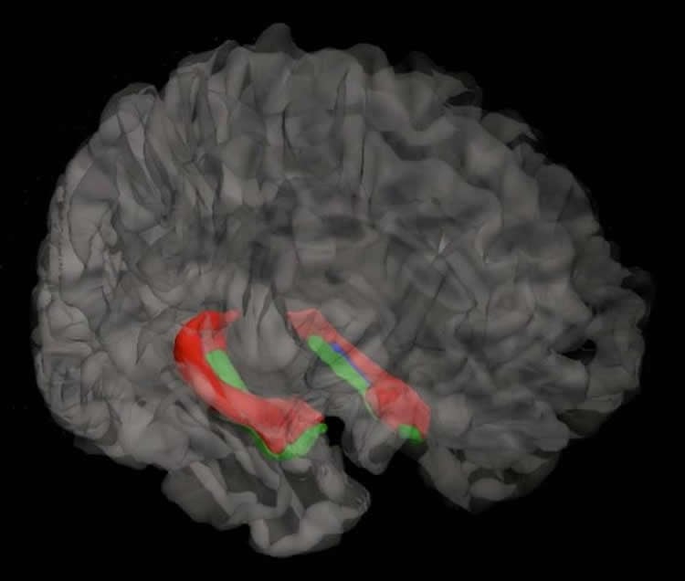
In order to compile this hippocampal atlas, researchers used MRI imagery from a sample of 25 healthy individuals. They then used their expertise in brain anatomy to label all the substructures composing the region, providing a model of an average, healthy hippocampus. The end result is analogous to a Google street view of this particular part of the brain. With this tool, researchers will be better able to assess the pathology of their patients by comparing their data to the atlas and will more clearly be able to locate the areas in need of surgical intervention.
A tool for brain diseases experts of all levels
“Our primary purpose was epilepsy. We wanted to be able to detect and identify different substructures in the hippocampus to enable us to be a lot more precise in our diagnosis and to pinpoint the affected region to better target treatments”, said Dr. Bernasconi. “With this new submillimetric dataset, made available through open science, we are not just sharing MRI images, we are also transferring anatomical knowledge and providing a statistical map that can be used by researchers and clinicians of different levels of expertise anywhere in the world.”
These tools hold promising therapeutic implications for epilepsy, but also for other neurological and psychiatric disorders such as Alzheimer’s disease, schizophrenia and depression. Crucially, the atlas provides researchers with a non-invasive way to assess the impact of therapies targeting this region of the brain and to thus develop better treatments to improve the quality of life for their patients.
This work conducted by Dr. Neda Bernasconi and collaborators at the Montreal Neurological Institute, was supported by the Canadian Institutes of Health Research.
Funding: The study was funded by Canadian Institutes of Health Research.
Source: Maya-Olivia Eyssen – McGill University
Image Source: The image is credited to Dr. Neda Bernasconi
Original Research: Full open access research for “Multi-contrast submillimetric 3 Tesla hippocampal subfield segmentation protocol and dataset” by Jessie Kulaga-Yoskovitz, Boris C. Bernhardt, Seok-Jun Hong, Tommaso Mansi, Kevin E. Liang, Andre J.W. van der Kouwe, Jonathan Smallwood, Andrea Bernasconi and Neda Bernasconi in Scientific Data. Published online November 10 2015 doi:10.1038/sdata.2015.59
Abstract
Multi-contrast submillimetric 3 Tesla hippocampal subfield segmentation protocol and dataset
The hippocampus is composed of distinct anatomical subregions that participate in multiple cognitive processes and are differentially affected in prevalent neurological and psychiatric conditions. Advances in high-field MRI allow for the non-invasive identification of hippocampal substructure. These approaches, however, demand time-consuming manual segmentation that relies heavily on anatomical expertise. Here, we share manual labels and associated high-resolution MRI data (MNI-HISUB25; submillimetric T1- and T2-weighted images, detailed sequence information, and stereotaxic probabilistic anatomical maps) based on 25 healthy subjects. Data were acquired on a widely available 3 Tesla MRI system using a 32 phased-array head coil. The protocol divided the hippocampal formation into three subregions: subicular complex, merged Cornu Ammonis 1, 2 and 3 (CA1-3) subfields, and CA4-dentate gyrus (CA4-DG). Segmentation was guided by consistent intensity and morphology characteristics of the densely myelinated molecular layer together with few geometry-based boundaries flexible to overall mesiotemporal anatomy, and achieved excellent intra-/inter-rater reliability (Dice index ≥90/87%). The dataset can inform neuroimaging assessments of the mesiotemporal lobe and help to develop segmentation algorithms relevant for basic and clinical neurosciences.
“Multi-contrast submillimetric 3 Tesla hippocampal subfield segmentation protocol and dataset” by Jessie Kulaga-Yoskovitz, Boris C. Bernhardt, Seok-Jun Hong, Tommaso Mansi, Kevin E. Liang, Andre J.W. van der Kouwe, Jonathan Smallwood, Andrea Bernasconi and Neda Bernasconi in Scientific Data. Published online November 10 2015 doi:10.1038/sdata.2015.59


