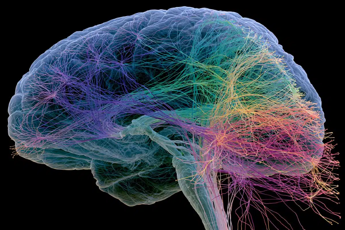
Summary: Researchers achieved a breakthrough in mapping the brain’s ‘dysfunctome’—key dysfunctional circuits linked to disorders like Parkinson’s, dystonia, OCD, and Tourette’s syndrome.
Utilizing deep brain stimulation (DBS) data from 261 patients worldwide, they pinpointed specific frontal cortex circuits crucial for symptom improvement. This novel map not only advances our understanding of neurologic and neuropsychiatric disorders but also opens new avenues for both surgical and non-invasive treatments like transcranial magnetic stimulation (TMS).
The study’s findings have already led to successful symptom alleviation in patients with severe OCD, demonstrating the potential for more personalized and effective therapies.
Key Facts:
- Comprehensive Brain Circuit Mapping: By analyzing data from 534 DBS electrodes, researchers identified vital networks in the brain’s frontal cortex associated with four distinct disorders.
- Human ‘Dysfunctome’: This work pioneers the concept of a ‘dysfunctome,’ a comprehensive map of brain circuits that become dysfunctional due to various neurologic and neuropsychiatric conditions.
- Potential for Personalized Therapy: The study’s insights into the dysfunctome allow for fine-tuning DBS and exploring non-invasive treatments like TMS, paving the way for tailored therapies based on specific brain circuit dysfunctions.
Source: Charite
When certain connections in the brain do not function correctly, disorders such as Parkinson’s disease, dystonia, obsessive-compulsive disorder (OCD), and Tourette’s syndrome may result. Targeted stimulation of specific areas in the brain can help alleviate symptoms.
To pinpoint the exact therapeutic target areas of the brain, a team led by researchers from Charité – Universitätsmedizin and Brigham and Women’s Hospital analyzed data from patients across the globe who had undergone implantation of tiny electrodes to stimulate the brain.
The result is a unique map of disrupted brain networks that has now been published in Nature Neuroscience.

Neurological and neuropsychiatric disorders present with a broad spectrum of different symptoms, from mood and information processing disorders to motor deficits. But they do have one thing in common: They are all attributable to malfunctioning connections between specific regions of the brain. In simplified terms, when brain circuits become dysfunctional, they may act as brakes on the brain functions that the circuit usually carries out.
Deep brain stimulation (DBS) targets these kinds of malfunctional circuits and can be instrumental in alleviating symptoms in various areas. In this neurosurgical approach, small electrodes are implanted into precisely defined target brain areas. The electrodes then chronically emit weak electrical pulses to the surrounding tissue.
The stimulation effects travel along neural pathways to more distant areas of the brain to unfold their full impact. But stimulation is not always successful. Even small discrepancies in electrode placement can lead to weaker effects of the treatment.
Which specific brain circuits need to be stimulated to achieve optimal outcomes when treating a range of symptoms was the question for an international team headed by neuroscientists Prof. Andreas Horn and Dr. Ningfei Li at Charité and Brigham and Women’s Hospital.
“Our goal was to better understand where in the brain possible ‘brakes’ can be released through neuromodulation to normalize the symptoms of Parkinson’s disease, for example,” says Ningfei Li.
Exploring a seeming paradox
The researchers’ work addresses a seeming paradox that has been known for a while in this field. A specific area of the basal ganglia called the subthalamic nucleus is considered an effective target for DBS to treat the symptoms of Parkinson’s disease and dystonia, which are both on the spectrum of movement disorders. Recently, the same region of the brain was also identified as a successful target for treating neuropsychiatric disorders such as OCD and tic disorders.
This raised the question of how such a small nucleus, only about one centimeter long, could be an effective target for symptoms of such different brain dysfunctions. To investigate this question, the team analyzed data from 534 DBS electrodes implanted in 261 patients across the globe.
Of this cohort, 70 patients were diagnosed with dystonia, 127 with Parkinson’s disease, 50 with OCD, and 14 with Tourette’s syndrome. Using software developed by the team, the researchers reconstructed the precise location of each electrode. Computer simulations were then used to map neural tracts that were activated in patients with optimal or suboptimal treatment outcomes.
Using these results, they were able to identify specific brain circuits that had become dysfunctional in each of the four disorders. They were associated with the relevant regions of the frontal part of the brain that play an important role in motor functions, impulse control, and information processing.
“The circuits we identified partially overlapped, which to us implies that the malfunctions reflected in the symptoms studied are not wholly independent from each other,” says Barbara Hollunder, a PhD fellow at the Einstein Center for Neurosciences at Charité and the first author of the study.
This means that as the first step, the researchers have succeeded in localizing the exact networks in the forebrain and midbrain that are crucial to treating Parkinson’s disease, dystonia, obsessive-compulsive disorder, and Tourette’s syndrome.
Applying this same approach across disorders with different symptoms gradually yields a map that denotes how specific brain circuits are associated with certain symptoms.
“By analogy to the terms ‘connectome,’ which describes a comprehensive map of all neural connections existing in the brain, and ‘genome,’ which is used for the full set of genetic information found in an organism, we have coined the term human ‘dysfunctome.’ One day, we hope the dysfunctome will describe the entirety of brain circuits that may typically become dysfunctional as a result of network disorders,” Hollunder explains.
Initial success with treatment in the course of the study
The researchers’ findings have already benefited the first few patients. Fine-tuning and precision electrode placement made it possible to alleviate the symptoms of severe, treatment-resistant OCD, for example.
“We plan to refine this technique and zero in even more precisely on dysfunctional brain circuits for specific symptoms. For example, we could isolate the circuits involved in obsessions or compulsions in OCD, or other comorbid symptoms commonly found in these patients, like depression and anxiety disorders, to individualize treatment further,” says Ningfei Li, looking to the future.
The researchers also believe that more than one region of the brain may be responsible for improvement of a given symptom. They suspect that neural networks themselves transmit the therapeutic effects, which can be modulated from various points in the brain.
This means the study provides not only valuable insights for targeted neurosurgical treatment, but may also inspire approaches for noninvasive neuromodulation such as transcranial magnetic stimulation (TMS), in which magnetic fields are used to stimulate certain areas of the brain from outside of the brain, without the need for surgery.
About this brain mapping research news
Author: Manuela Zingl
Source: Charite
Contact: Manuela Zingl – Charite
Image: The image is credited to Neuroscience News
Original Research: Open access.
“Mapping Dysfunctional Circuits in the Frontal Cortex Using Deep Brain Stimulation” by Barbara Hollunder et al. Nature Neuroscience
Abstract
Mapping Dysfunctional Circuits in the Frontal Cortex Using Deep Brain Stimulation
Frontal circuits play a critical role in motor, cognitive and affective processing, and their dysfunction may result in a variety of brain disorders. However, exactly which frontal domains mediate which (dys)functions remains largely elusive.
We studied 534 deep brain stimulation electrodes implanted to treat four different brain disorders. By analyzing which connections were modulated for optimal therapeutic response across these disorders, we segregated the frontal cortex into circuits that had become dysfunctional in each of them.
Dysfunctional circuits were topographically arranged from occipital to frontal, ranging from interconnections with sensorimotor cortices in dystonia, the primary motor cortex in Tourette’s syndrome, the supplementary motor area in Parkinson’s disease, to ventromedial prefrontal and anterior cingulate cortices in obsessive-compulsive disorder.
Our findings highlight the integration of deep brain stimulation with brain connectomics as a powerful tool to explore couplings between brain structure and functional impairments in the human brain.