State-of-the-art imaging technology for biomarker development is explored in a supplement to Journal of Alzheimer’s Disease.
Alzheimer’s disease (AD) is one of the most common brain disorders, with an estimated 35 million people affected worldwide. In the last decade, research has advanced our understanding of how AD affects the brain. However, diagnosis continues to rely primarily on neuropsychological tests which can only detect the disease after clinical symptoms begin. In a supplement to the Journal of Alzheimer’s Disease, investigators report on the development of imaging-based biomarkers that will have an impact on diagnosis before the disease process is set in motion.
“There is an urgent need for the development of reliable diagnostic biomarkers that can detect AD pathology at an incipient phase,” says Guest Editor Dr. Pravat Mandal, Adjunct Associate Professor of the Department of Radiology, Johns Hopkins Medicine, Baltimore, MD and Additional Professor, National Brain Research Center, India. “This special issue focuses on the latest strides made in identifying diagnostic biomarkers using state-of-the-art imaging modalities.”
The issue looks at the application of various magnetic resonance imaging (MRI) technologies for diagnosing AD and monitoring the progression of the disease. For example, Brian T. Gold and colleagues report on the use of diffusion tension imaging (DTI) to identify changes in the white matter of patients with amnestic mild cognitive impairment (MCI), an early symptom of AD. Charles D. Smith and colleagues describe MRI-based detection of key structural alterations in cognitively normal subjects that can serve as a predictor of memory impairment.

While MRI provides information about the anatomy of the brain, functional MRI (fMRI) provides crucial information about the regions involved during specific tasks. The issue explores how fMRI can detect changes in functional activation and connectivity in AD patients. Monitoring alterations in functional brain activity related to visual processing deficits in AD has immense potential as an early diagnostic biomarker, according to a review by Dr. Mandal and colleagues. By using fMRI to study normal older individuals, patients with MCI, and those with AD as they perform cognitive tasks and at rest, Jasmeer P. Chhatwal and Reisa Sperling reveal the functional alterations associated with healthy aging as well as MCI and AD.
Magnetic resonance spectroscopy (MRS) is a powerful non-invasive imaging technique that can provide crucial information about neurochemical changes in AD. Dr. Mandal and colleagues report that the identification of neurochemical changes in the brains of MCI and AD patients may provide a “signature” of early AD pathology, and may aid in diagnosing patients who are moving from MCI to more advanced AD.
Imaging is also used to examine the molecular and therapeutic effects of potential AD treatments. Liam Zaidel and colleagues found that patients treated with donepezil for mild cognitive symptoms had a significant increase in interhemispheric functional connectivity of the left and right dorsolateral prefrontal cortices. Giulia Liberati and colleagues describe a non-invasive brain-computer interface that can detect a patient’s emotional and cognitive state. It could provide vital information on the effect of clinical drugs on brain function and cognition in patients with AD.
The issue also includes a thought-provoking argument from Edo Richard and colleagues calling for a paradigm shift in dementia research and biomarker development. Current biomarker research focuses on correlates of plaques and tangles, which are poor markers in older dementia subjects. The authors suggest that the acknowledgement that dementia in older subjects is different from dementia at a young age will lead to new approaches in biomarker development and research.
“Research is needed to understand which molecular, structural, and functional changes are causally related to the onset of AD,” says Dr. Mandal. “This special issue aims to conceptualize more effective and reliable biomarkers for AD.”
George Perry, PhD, Editor-in-Chief, Journal of Alzheimer’s Disease, and Dean and Professor, College of Sciences, University of Texas at San Antonio, says, “The development of biomarkers to aid in early detection of the onset of AD is critical. This special issue will spur research into multi-model imaging based biomarker development for AD.”
Notes about this Alzheimer’s disease research
Contacts: George Perry, PhD – Editor-in-Chief at the Journal of Alzheimer’s Disease, Dean and Professor of Biology at The University of Texas at San Antonio
Daphne Watrin – IOS Press
Source: IOS Press press release
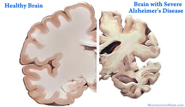
Image Source: This NeuroscienceNews.com Alzheimer’s disease brain image is in the public domain. The original image is at the NIH Medline Plus page for the “7 Warning Signs of Alzheimer’s”. Feel free to use our image.
Original Research: Supplement (in press) “Biomarkers for Alzheimer’s Disease Using Multi-Model Imaging Research” guest edited by Pravat K. Mandal in Journal of Alzheimer’s Disease 2012 Volume 31 Supplement 3


