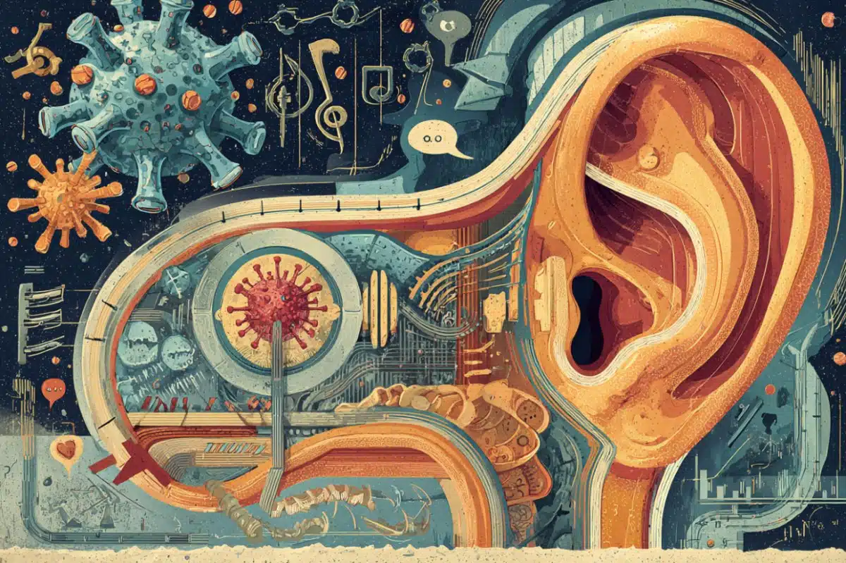
Summary: Compared to the virus that caused the SARS epidemic in the early 2000s, SARS-CoV-2, the virus responsible for COVID-19, has evolved new strategies to bind to cell receptors, resulting in tighter binding. A new study identifies key mutations that potentially enabled the animal-to-human transmission of SARS-CoV-2. The study mapped out important binding sites on SARS-CoV-2 for antibody drugs to act on. If new drugs can bind to the sites on SARS-CoV-2, they will block the virus out of cells.
Source: University of Minnesota
Coronaviruses infect humans by binding to specific proteins, known as receptors, on human cell surfaces. Researchers from the University of Minnesota, led by Professor Fang Li in the Department of Veterinary and Biomedical Sciences at the College of Veterinary Medicine, recently broke new ground in understanding how SARS-CoV-2, the virus that causes COVID-19, binds to its human receptor. Their findings were recently published in the journal Nature. This knowledge not only facilitates a better understanding of the infectivity of the virus, but also sheds light on its animal origin and provides guidance on vaccine and antiviral drug designs.
Several technologies, all with intrinsic limitations, have recently provided some information on how SARS-CoV-2 binds to its receptor. Li’s study is the first to use x-ray crystallography, the “gold-standard” method of structural biology at atomic resolution, to map out the 3D structure of a protein on SARS-CoV-2 that binds to its human receptor. This 3D imagery reveals much information about how the two proteins connect.
More specifically, the 3D structure shows that:
- compared to the virus that caused the 2002-2003 SARS outbreak, the new coronavirus has evolved new strategies to bind to its human receptor, resulting in tighter binding of the receptor; and
- two related coronaviruses, one found in bats and one found in pangolins, can bind directly to the same human receptor as SARS-CoV-2, suggesting that SARS-CoV-2 originates from bats, either directly or with pangolins as an intermediate host. To infect humans, however, the bat or pangolin coronavirus needed to undergo mutations to gain more efficient usage of the human receptor.
The study has identified key mutations that potentially enabled the animal-to-human transmission of SARS-CoV-2.
With the 3D structure in hand, the study has mapped out the important binding sites on SARS-CoV-2 for antibody drugs to act on, providing a blueprint for developing new antibody drugs that specifically target those sites. If a new antibody drug can bind to those sites on the virus more strongly and frequently than the receptor, it will block the virus out of cells, making it a potentially effective treatment for viral infections. Those sites are also valuable for vaccine designs, as vaccines containing those sites can induce the production of antibodies in the human body, which can prevent future viral infections.

Next, the research team plans to use structural information from this study to develop antibody drugs and vaccines that specifically target the binding of SARS-CoV-2 to its human receptor.
Other contributors to the study are Jian Shang, Gang Ye, Yushun Wan, Chuming Luo, Qibin Geng, Ashley Auerbach from Department of Veterinary and Biomedical Sciences, and Ke Shi, Hideki Aihara from Department of Biochemistry, Molecular Biology and Biophysics in the College of Biological Sciences and the Medical School. The study was funded by the National Institute of Health.
Source:
University of Minnesota
Media Contacts:
University Public Relations – University of Minnesota
Image Source:
The image is adapted from the University of Minnesota news release.
Original Research: Closed access
“Structural basis of receptor recognition by SARS-CoV-2”. Jian Shang, Gang Ye, Ke Shi, Yushun Wan, Chuming Luo, Hideki Aihara, Qibin Geng, Ashley Auerbach & Fang Li.
Nature doi:10.1038/s41586-020-2179-y.
Abstract
Structural basis of receptor recognition by SARS-CoV-2
A novel SARS-like coronavirus (SARS-CoV-2) recently emerged and is rapidly spreading in humans1,2. A key to tackling this epidemic is to understand the virus’s receptor recognition mechanism, which regulates its infectivity, pathogenesis and host range. SARS-CoV-2 and SARS-CoV recognize the same receptor – human ACE2 (hACE2)3,4. Here we determined the crystal structure of the SARS-CoV-2 receptor-binding domain (RBD) (engineered to facilitate crystallization) in complex with hACE2. Compared with the SARS-CoV RBD, a hACE2-binding ridge in SARS-CoV-2 RBD takes a more compact conformation; moreover, several residue changes in SARS-CoV-2 RBD stabilize two virus-binding hotspots at the RBD/hACE2 interface. These structural features of SARS-CoV-2 RBD enhance its hACE2-binding affinity. Additionally, we show that RaTG13, a bat coronavirus closely related to SARS-CoV-2, also uses hACE2 as its receptor. The differences among SARS-CoV-2, SARS-CoV and RaTG13 in hACE2 recognition shed light on potential animal-to-human transmission of SARS-CoV-2. This study provides guidance for intervention strategies targeting receptor recognition by SARS-CoV-2.