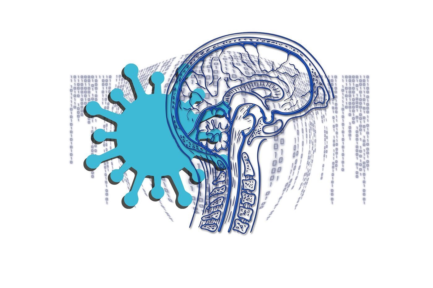
Summary: Some coronavirus patients exhibit clinical and neurochemical signs of brain injury associated with the viral infection. COVID-19 patients who required ventilation had increased plasma NfL levels. Higher NfL concentration levels were linked to the severity of the infection.
Source: University of Gothenburg
Certain patients who receive hospital care for coronavirus infection (COVID-19) exhibit clinical and neurochemical signs of brain injury, a University of Gothenburg study shows. In even moderate COVID-19 cases, finding and measuring a blood-based biomarker for brain damage proved to be possible.
Some people infected with the coronavirus SARS-CoV-2 get only mild, cold-like symptoms, while others become severely ill and require hospital treatment. Among the latter, it has become clear that the patients sometimes show obvious signs of the brain not functioning as it should. These cases are not common, but do occur.
In a project at Sahlgrenska Academy, University of Gothenburg, blood samples were taken from 47 patients with mild, moderate and severe COVID-19 in the course of their hospital stay. These samples were analyzed by means of highly sensitive biomarkers for brain injury. The results were compared with those from a healthy control group comprising 33 people matched by age and sex.
Now that the research is being presented in the journal Neurology, it is evident that an increase in one of the biomarkers took place even with moderate COVID-19 — that is, in patients admitted to hospital but not in need of ventilator support. This marker, known as GFAP (glial fibrillary acidic protein), is normally present in astrocytes, a star-shaped neuron-supportive cell type in the brain, but leaks out in the event of astrocytic injury or overactivation.
The second biomarker investigated was NfL (neurofilament light chain protein), which is normally to be found inside the brain’s neuronal outgrowths, which it serves to stabilize, but which leaks out into the blood if they are damaged. Elevated plasma NfL concentrations were found in most of the patients who required ventilator treatment, and there was a marked correlation between how much they rose and the severity of the disease.
“The increase in NfL levels, in particular, over time is greater than we’ve seen previously in studies connected with intensive care, and this suggests that COVID-19 can in fact directly bring about a brain injury. Whether it’s the virus or the immune system that’s causing this is unclear at present, and more research is needed,” says Henrik Zetterberg, Professor of Neurochemistry, whose research team at Sahlgrenska Academy performed the measurements.
Magnus Gisslén, Professor of Infectious Diseases at Sahlgrenska Academy and chief physician at the Department of Infectious Diseases, Sahlgrenska University Hospital, leads the Academy’s clinical research on COVID-19.
In his view, blood tests for biomarkers associated with brain injury could be used for monitoring patients with moderate to severe COVID-19, to reduce the risk of brain injury.
“It would be highly interesting to see whether the NfL increase can be slowed down with new therapies, such as the new dexamethasone treatment that’s now been proposed,” Gisslén says.
About this neuroscience research article
Source:
University of Gothenburg
Media Contacts:
Henrik Zetterberg – University of Gothenburg
Image Source:
The image is in the public domain.
Original Research: Closed access
“Neurochemical evidence of astrocytic and neuronal injury commonly found in COVID-19”. by Nelly Kanberg, Nicholas J. Ashton, Lars-Magnus Andersson, Aylin Yilmaz, Magnus Lindh, Staffan Nilsson, Richard W Price, Kaj Blennow, Henrik Zetterberg, Magnus Gisslén.
Neurology doi:10.1212/WNL.0000000000010111
Abstract
Neurochemical evidence of astrocytic and neuronal injury commonly found in COVID-19
Objective: To test the hypothesis that COVID-19 has an impact on the CNS by measuring plasma biomarkers of CNS injury.
Methods: We recruited 47 patients with mild (n=20), moderate (n=9) or severe (n=18) COVID-19 and measured two plasma biomarkers of CNS injury by Single molecule array (Simoa): neurofilament light chain protein (NfL) (a marker of intra-axonal neuronal injury) and glial fibrillary acidic protein (GFAp) (a marker of astrocytic activation/injury) in samples collected at presentation and again in a subset after a mean of 11.4 days. Cross-sectional results were compared with 33 age-matched controls derived from an independent cohort.
Results: The patients with severe COVID-19 had higher plasma concentrations of GFAp (p=0.001) and NfL (p<0.001) than controls, while GFAp was also increased in patients with moderate disease (p=0.03). In severe patients an early peak in plasma GFAp decreased upon follow-up (p<0.01) while NfL showed a sustained increase from first to last follow-up (p<0.01), perhaps reflecting a sequence of early astrocytic response and more delayed axonal injury.
Conclusion: We show neurochemical evidence of neuronal injury and glial activation in patients with moderate and severe COVID-19. Further studies are needed to clarify the frequency and nature of COVID-19-related CNS damage, and its relation to both clinically-defined CNS events such as hypoxic and ischemic events and to mechanisms more closely linked to systemic SARS-CoV-2 infection and consequent immune activation, and also to evaluate the clinical utility of monitoring plasma NfL and GFAp in management of this group of patients.


