Summary: Our brains demand a huge amount of energy, yet they allocate blood with remarkable efficiency. Researchers have uncovered how endothelial cells lining blood vessels act as a rapid “signaling highway,” using gap junctions to direct blood precisely where brain activity is highest.
This mechanism allows the brain to prioritize its limited energy, and its breakdown may underlie neurodegenerative diseases. The findings could improve understanding of fMRI scans and pave the way for new therapies for brain disorders.
Key Facts:
- Signaling Highway: Endothelial cells use gap junctions to rapidly communicate blood needs across brain regions.
- Energy Efficiency: This mechanism helps the brain allocate blood to active regions despite its high energy demands.
- Disease Insight: Dysfunction of this system may contribute to neurodegeneration, offering a new therapeutic target.
Source: Harvard
All day long, our brains carry out complicated and energy-intensive tasks such as remembering, solving problems, and making decisions.
To supply the energy these tasks require while conserving this precious fuel, the brain has evolved a system that allows it to quickly and efficiently send blood only to the areas that need it most in any given moment. This system is essential to brain function and overall health, yet how it works has remained somewhat of a mystery.
Now, a team led by researchers at Harvard Medical School has uncovered new details of how the brain moves blood to active areas in real time.
Their findings are published July 16 in Cell.
In experiments in mice, the team discovered that the brain uses specialized channels in the lining of its blood vessels to communicate where blood is needed.
“This work helps us understand how you can get that super-important blood supply to the correct areas of the brain on a time scale that is useful,” said co-lead author Luke Kaplan, a research fellow in neurobiology in the Blavatnik Institute at HMS.
If confirmed in additional studies in animals and humans, the findings could be used to better understand findings on brain imaging tests such as functional MRI (fMRI). The insights may also advance understanding of neurodegenerative diseases, in which this communication system often breaks down, leading to cognitive problems.
A long-standing mechanistic puzzle
In the late 1800s, Italian physician Angelo Mosso observed something intriguing in a patient with a skull defect that left an area of his brain exposed: When the patient got angry, parts of the exposed area instantly swelled with blood — hinting at a connection between brain activity and blood flow.
A century later, this connection became the basis of fMRI, a type of brain scan that measures blood flow to different regions as a proxy for neural activity while individuals perform various tasks.
The brain is one of the body’s most energy-demanding organs, accounting for 2 percent of the body’s weight but consuming 20 percent of its total energy. To stay within budget, the brain must be highly efficient: It rapidly directs blood flow to the regions that need it the most, explained senior author Chenghua Gu, professor of neurobiology at HMS.
While the brain’s proportion of energy consumption varies across species depending on the organ’s complexity, Gu said, the brain is an energy-hungry organ in all mammals.
“There’s this an elegant evolutionary mechanism that’s distributing blood flow on demand throughout the brain, but we don’t understand how it works,” said co-lead author Trevor Krolak, a doctoral student in the Gu lab.
This blood-allocation process deteriorates in neurodegeneration, Kaplan noted, and understanding its inner workings could lead to new insights.
“If we don’t know how this system operates on a biological level, we won’t know how to fix it when it goes wrong,” he said.
A cellular signaling highway revealed
To understand what happens on a molecular level as different brain regions become active, the researchers performed a series of experiments in mice. Their analysis showed the brain rapidly communicates the need for more blood in a particular area via endothelial cells that line blood vessels in the brain.
Furthermore, the researchers observed, the endothelial cells communicated so quickly and efficiently via so-called gap junctions — tiny channels that physically connect neighboring cells. The team also identified two genes pivotal to the function of this communication pathway.
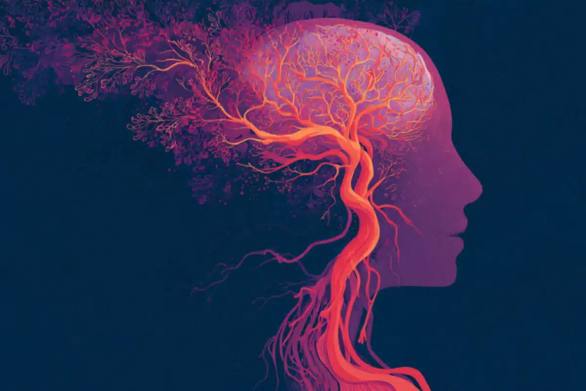
“The brain essentially uses the inner lining of its blood vessels as a wide-ranging and coordinated signaling highway,” Krolak said.
In doing so, the brain can communicate which blood vessels need to dilate or contract in concert to move blood to the right areas at the right time.
“These findings help us understand how the brain delivers blood flow to exactly where it is needed, allowing it to utilize its limited energy supply in an efficient way,” Gu said.
Other types of cells are also connected by gap junctions, and mutations in gap junction genes have been implicated in heart conditions, deafness, and other diseases. Thus, the researchers noted, the new findings open the door for more research on how cells throughout the body use these connections to communicate.
Because brain vasculature is highly conserved in mammals, Gu said, the same system likely operates in humans. If the findings, indeed, translate to humans, the researchers think they could improve interpretation of fMRI, which relies on the link between blood flow and neural activity.
A growing body of evidence suggests that regulation of blood supply is important for brain health — and begins to break down in certain neurodegenerative diseases, the researchers said. The team hopes that their findings can shed light on the changes that occur in the brain during neurodegeneration.
“Now that we’ve figured out the mechanism” Gu said, “we want to apply our knowledge to understanding disease and developing therapies.”
Authorship, funding, disclosures
Additional authors on the paper include Kathleen Navas, Lujing Chen, Austin Birmingham, Daniel Ryvkin, Victoria Izsa, Megan Powell, Zhuhao Wu, and Benjamin Deverman.
Funding: The study was funded by a National Science Foundation Graduate Research Fellowship, an HMS Mahoney Postdoctoral Fellowship, an HMS Lefler Postdoctoral Fellowship, the National Institutes of Health (RF1MH128969; HL153261; R35NS116820), the National Institute of Neurological Disorders and Stroke (UG3NS111689), the National Institute of Mental Health (UG3MH120096), the Stanley Center for Psychiatric Research, and a Fidelity Biosciences Research Initiative. Gu is an investigator of the Howard Hughes Medical Institute.
Deverman is listed as an inventor on a patent application (US20240325568A1) for production and use of the AAV-BI30 vector. Deverman is a scientific founder and scientific advisor of Apertura Gene Therapy, received research funding from Apertura Gene Therapy, and is on the scientific advisory board of Tevard Biosciences.
About this neuroscience research news
Author: Ekaterina Pesheva
Source: Harvard
Contact: Ekaterina Pesheva – Harvard
Image: The image is credited to Neuroscience News
Original Research: Open access.
“Brain Endothelial Gap Junction Coupling Enables Rapid Vasodilation Propagation During Neurovascular Coupling” by Luke Kaplan et al. Cell
Abstract
Brain Endothelial Gap Junction Coupling Enables Rapid Vasodilation Propagation During Neurovascular Coupling
To meet the brain’s moment-to-moment energy demand, neural activation rapidly increases local blood flow. This process, known as neurovascular coupling, involves rapid, coordinated vasodilation of the brain’s arterial network.
Here, we demonstrate that endothelial gap junction coupling enables long-range propagation of vasodilation signals through the vasculature during neurovascular coupling.
The molecular composition of these gap junctions is zonated along the arterio-venous axis, with arteries being the most strongly coupled segment.
Using optogenetics and visual stimuli in awake mice, we found that acute, arterial endothelial cell type-specific deletion of Cx37 and Cx40 abolishes arterial gap junction coupling and results in impaired vasodilation.
Specifically, we demonstrated that arterial endothelial gap junction coupling determines both the speed and the spatial extent of vasodilation propagation elicited by neural activity.
These findings indicate that endothelial gap junctions serve as a signaling highway for neurovascular coupling, enabling flexible and efficient distribution of limited energetic resources.