

Summary: Scientists have created a groundbreaking spatial cell atlas of the human limb, capturing the intricate process of human limb development. This work, part of the Human Cell Atlas initiative, marks a significant advancement in understanding the rapid and complex formation of human limbs.
The study provides a detailed map of the cellular landscape during early limb formation, offering insights into congenital limb syndromes. This research not only deepens our understanding of human anatomy but also opens avenues for diagnosing and treating various limb-related disorders.
Key Facts:
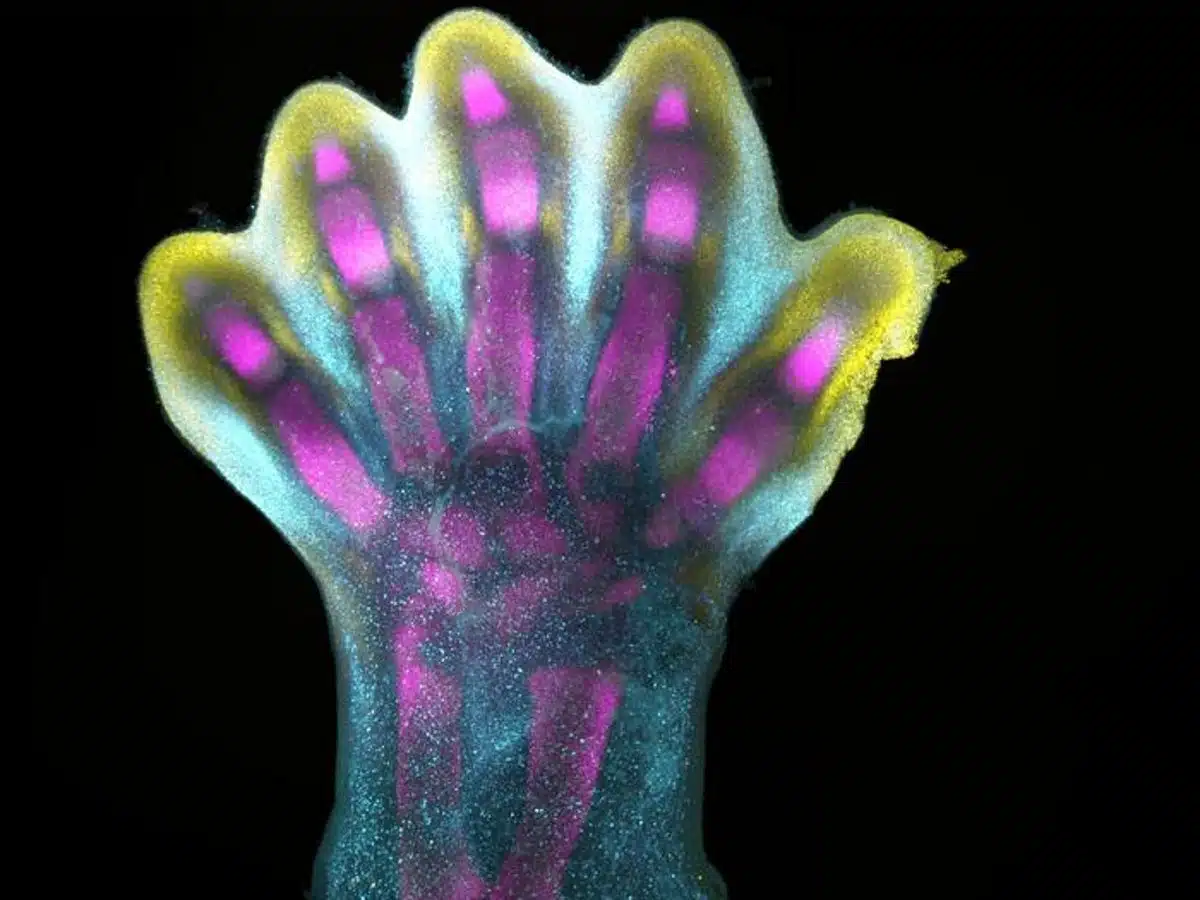
- The atlas showcases how human fingers and toes form from a larger foundational bud, with intervening cells receding to reveal the digits.
- This study is a part of the international Human Cell Atlas initiative, aiming to map every cell type in the human body to revolutionize our understanding of health and disease.
- The atlas uncovers new links between developmental cells and congenital limb syndromes, such as brachydactyly (short fingers) and polysyndactyly (extra digits).
Source: Wellcome Sanger Institute
Human fingers and toes do not grow outward; instead, they form from within a larger foundational bud, as intervening cells recede to reveal the digits beneath. This is among many processes captured for the first time as scientists unveil a spatial cell atlas of the entire developing human limb, resolved in space and time.
Researchers at the Wellcome Sanger Institute, Sun Yat-sen University, EMBL’s European Bioinformatics Institute and collaborators applied cutting-edge single-cell and spatial technologies to create an atlas characterising the cellular landscape of the early human limb, pinpointing the exact location of cells.
This study is part of the international Human Cell Atlas initiative to map every cell type in the human body, to transform understanding of health and disease.
The atlas, published today (6 December) in Nature, provides an openly available resource that captures the intricate processes governing the limbs’ rapid development during the early stages of limb formation.
The atlas also uncovers new links between developmental cells and some congenital limb syndromes, such as short fingers and extra digits.
Limbs are known to initially emerge as undifferentiated cell pouches on the sides of the body, without a specific shape or function. However after 8 weeks of development, they are well differentiated, anatomically complex and immediately recognisable as limbs, complete with fingers and toes.
This requires a very rapid and precise orchestration of cells. Any small disturbances to this process can have a downstream effect, which is why variations in the limbs are among the most frequently reported syndromes at birth, affecting approximately one in 500 births globally.
While limb development has been extensively studied in mouse and chick models, the extent to which they mirror the human situation remained unclear. However, advances in technology now enable researchers to explore the early stages of human limb formation.
In this new study, scientists from the Wellcome Sanger Institute, Sun Yat-sen University, and their collaborators analysed tissues between 5 and 9 weeks of development. This allowed them to trace specific gene expression programs, activated at certain times and in specific areas, which shape the forming limbs.
Special staining of the tissue revealed clearly how cell populations differentially arrange themselves into patterns of the forming digits.
As part of the study, researchers demonstrated that certain gene patterns have implications for how the hands and feet form, identifying certain genes, which when disrupted, are associated with specific limb syndromes like brachydactyly – short fingers – and polysyndactyly – extra fingers or toes.
The team were also able to confirm that many aspects of limb development are shared between humans and mice.
Overall, these findings not only provide an in-depth characterisation of limb development in humans but also critical insights that could impact the diagnosis and treatment of congenital limb syndromes.
Professor Hongbo Zhang, senior author of the study from Sun Yat-sen University, Guangzhou, said: “Decades of studying model organisms established the basis for our understanding of vertebrate limb development. However, characterising this in humans has been elusive until now, and we couldn’t assume the relevance of mouse models for human development.
“What we reveal is a highly complex and precisely regulated process. It is like watching a sculptor at work, chiselling away at a block of marble to reveal a masterpiece. In this case, nature is the sculptor, and the result is the incredible complexity of our fingers and toes.”
Dr Sarah Teichmann, senior author of the study from the Wellcome Sanger Institute, and co-founder of the Human Cell Atlas, said: “For the first time, we have been able to capture the remarkable process of limb development down to single cell resolution in space and time.
“Our work in the Human Cell Atlas is deepening our understanding of how anatomically complex structures form, helping us uncover the genetic and cellular processes behind healthy human development, with many implications for research and healthcare.
“For instance, we discovered novel roles of key genes MSC and PITX1 that may regulate muscle stem cells. This could offer potential for treating muscle-related disorders or injuries.”
About this cell mapping and developmental neuroscience research news
Author: Jelena Pupavac
Source: Wellcome Sanger Institute
Contact: Jelena Pupavac – Wellcome Sanger Institute
Image: The image is credited to the researchers/Nature
Original Research: Open access.
“A human embryonic limb cell atlas resolved in space and time” by Sarah Teichmann et al. Nature
Abstract
A human embryonic limb cell atlas resolved in space and time
Human limbs emerge during the fourth post-conception week as mesenchymal buds, which develop into fully formed limbs over the subsequent months. This process is orchestrated by numerous temporally and spatially restricted gene expression programmes, making congenital alterations in phenotype common.
Decades of work with model organisms have defined the fundamental mechanisms underlying vertebrate limb development, but an in-depth characterization of this process in humans has yet to be performed.
Here we detail human embryonic limb development across space and time using single-cell and spatial transcriptomics. We demonstrate extensive diversification of cells from a few multipotent progenitors to myriad differentiated cell states, including several novel cell populations.
We uncover two waves of human muscle development, each characterized by different cell states regulated by separate gene expression programmes, and identify musculin (MSC) as a key transcriptional repressor maintaining muscle stem cell identity.
Through assembly of multiple anatomically continuous spatial transcriptomic samples using VisiumStitcher, we map cells across a sagittal section of a whole fetal hindlimb. We reveal a clear anatomical segregation between genes linked to brachydactyly and polysyndactyly, and uncover transcriptionally and spatially distinct populations of the mesenchyme in the autopod.
Finally, we perform single-cell RNA sequencing on mouse embryonic limbs to facilitate cross-species developmental comparison, finding substantial homology between the two species.


