

Summary: Study examines the interactions between stem cells and neurons in both tissue regeneration and the development of cancerous tumors.
Source: University of Zurich
Stem cells can generate a variety of specific tissues and are increasingly used for clinical applications such as the replacement of bone or cartilage. However, stem cells are also present in cancerous tissues and are involved in cancer progression and metastasis. Nerves are fundamental for regulating the physiological and regenerative processes involving stem cells. However, little is known about the interactions between stem cells and neurons in regenerating tissues and in cancers.
Comparing stem cell types in tissue regeneration
A team of researchers led by Thimios Mitsiadis, professor at the Institute of Oral Biology of the University of Zurich, has now published two studies that elucidate how stem cells promote neuronal growth in tissue regeneration and in cancer progression. The first study compared the interaction of neurons with two different human stem cell populations, namely dental pulp stem cells and bone marrow stem cells. Both can differentiate into various cell types such as bone, cartilage and fat cells. Human bone marrow stem cells are isolated from skeletal bones and are the gold standard for bone regenerative approaches. Extracted teeth are the source of dental pulp stem cells, which represent a promising alternative.
Dental stem cells are highly innervated
Using the “organ-on-a-chip” technology, which relies on small three-dimensional devices mimicking the basic functions of human organs and tissues, the researchers demonstrated that both types of stem cells promoted neuronal growth. The dental pulp stem cells, however, yielded better results compared to bone marrow stem cells: They induced more elongated neurons, formed dense neuronal networks and established close contacts with nerves.
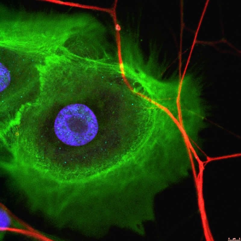
“Dental stem cells produce specific molecules that are fundamental for the growth and attraction of neurons. Therefore, stem cells are abundantly innervated,” says Mitsiadis. The formation of such extended networks and the establishment of numerous contacts suggest that dental stem cells create functional connections with nerves of the face. “Therefore, these cells could represent an attractive choice for the regeneration of functional, properly innervated facial tissues,” adds co-author and junior group leader Pierfrancesco Pagella.
Cancer stem cells also recruit neurons
In the second study, the researchers examined the interaction between nerves and cancer stem cells found in ameloblastoma, an aggressive tumour of the mouth. They first demonstrated that ameloblastomas have stem cell properties and are innervated by facial neurons. When ameloblastoma cells were isolated and placed in the “organ-on-a-chip” devices, they retained not only their stem cell properties but also attracted nerves and established contact with them.
“It appears that nerves are fundamental for the survival and function of cancer stem cells,” explains Pagella. “These results create new possibilities for cancer treatment using drugs that modify the communication between neurons and cancer stem cells. We hope this opens unforeseen paths towards effective therapies against cancer,” adds Mitsiadis. “The combination of advanced molecular and imaging tools and “organ-on-a-chip” technology offers an exciting opportunity to reveal the hidden functions of neurons and their interactions with various stem cell types, in both healthy and pathological conditions.”
Source:
University of Zurich
Media Contacts:
Thimios Mitsiadis – University of Zurich
Image Source:
The image is credited to the Institute of Oral Biology, University of Zurich.
Original Research: Open access
“Ameloblastomas Exhibit Stem Cell Potential, Possess Neurotrophic Properties, and Establish Connections with Trigeminal Neurons”. Thimios Mitsiadis et al.
Cells doi:10.3390/cells9030644.
Abstract
Ameloblastomas Exhibit Stem Cell Potential, Possess Neurotrophic Properties, and Establish Connections with Trigeminal Neurons
Ameloblastomas are locally invasive and aggressive odontogenic tumors treated via surgical resection, which results in facial deformity and significant morbidity. Few studies have addressed the cellular and molecular events of ameloblastoma onset and progression, thus hampering the development of non-invasive therapeutic approaches. Tumorigenesis is driven by a plethora of factors, among which innervation has been long neglected. Recent findings have shown that innervation directly promotes tumor progression. On this basis, we investigated the molecular characteristics and neurotrophic properties of human ameloblastomas. Our results showed that ameloblastomas express dental epithelial stem cell markers, as well as components of the Notch signaling pathway, indicating persistence of stemness. We demonstrated that ameloblastomas express classical stem cell markers, exhibit stem cell potential, and form spheres. These tumors express also molecules of the Notch signaling pathway, fundamental for stem cells and their fate. Additionally, we showed that ameloblastomas express the neurotrophic factors NGF and BDNF, as well as their receptors TRKA, TRKB, and P75/NGFR, which are responsible for their innervation by trigeminal axons in vivo. In vitro studies using microfluidic devices showed that ameloblastoma cells attract and form connections with these nerves. Innervation of ameloblastomas might play a key role in the onset of this malignancy and might represent a promising target for non-invasive pharmacological interventions.


