Summary: Neurofeedback training helps reinforce brain patterns associated with motor function while participants complete imaginary motor tasks. The findings may help in the rehabilitation for stroke and other disorders that compromise motor function.
Source: D’Or Institute for Research and Education
Less than one hour of brain training with neurofeedback leads to a strengthening of neural connections and communication among brain areas. This is the main finding of a new study conducted at D’Or Institute for Research and Education (IDOR), published in Neuroimage. According to the authors, the study may pave the way for the optimization and development of therapeutic approaches against stroke and Parkinson’s, for example.
“We knew that the brain has an amazing ability to adapt itself, but we were not sure that we could observe these changes so quickly. Understanding of how we can impact on brain wiring and functioning is the key to treat neurological disorders”, says Theo Marins, a biomedical scientist from IDOR and the Ph.D. responsible for the study.
Neurofeedback has been considered a promising way to regulate dysfunctional brain areas associated with disorders, such as chronic pain and depression, for example. With this technique, the magnetic resonance equipment helps individuals have access to their own brain activity in real time and quickly gain control over it.
Thirty-six healthy subjects participated in the study in which the goal was to increase the activity of brain regions involved in hand movements. However, instead of actually move their hand, participants were asked to only imagine the movement, in total rest. Nineteen of them received the real brain training and the remaining seventeen were trained with placebo neurofeedback, for comparison purposes. Immediately before and after the brain training, which lasted around 30 minutes, their neural networks were scanned in order to investigate the impact of the neurofeedback (or placebo) on brain wiring and communication, also known as structural and functional connectivity, respectively.

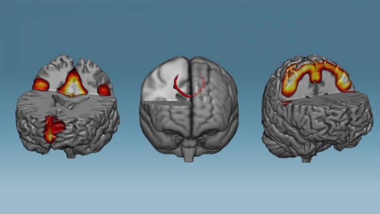
The results show that the corpus callosum – the major cerebral bridge that connects the right and left hemispheres – exhibited increased integrity, and the neural network controlling the movements of the body became strengthened. It seems that the whole system became more robust. Likewise, the training also had a positive impact on the default mode network, a brain network which is impaired after stroke, Parkinson’s and depression, for example. These changes were not observed in the control group.
“We showed that the neurofeedback can be considered a powerful tool to induce brain changes at record speed. Now, our goal is to develop new studies to test whether patients with neurological disorders can also benefit from it”, concludes Fernanda Tovar Moll, president of IDOR and leader of the study.
The study was conducted in collaboration with Federal University of Rio de Janeiro (UFRJ) and Augusto Motta University (Unisuam) and was part of the Ph.D. thesis of Theo Marins.
Source:
D’Or Institute for Research and Education
Media Contacts:
Helen Santoro – D’Or Institute for Research and Education
Image Source:
The image is credited to D’Or Institute for Research and Education (IDOR).
Original Research: Closed access
“Structural and functional connectivity changes in response to short-term neurofeedback training with motor imagery”
T. Marins, E. Rodrigues, T. Bortolini, Bruno Melo, J. Moll, F. Tovar-Moll. NeuroImage 03 APR 2019 doi:10.1016/j.neuroimage.2019.03.027
Abstract
Structural and functional connectivity changes in response to short-term neurofeedback training with motor imagery
Recent findings have been challenging current understanding about how fast the human brain change its structural and functional connections in response to training. One powerful way to deepen the inner workings of human brain plasticity is using neurofeedback (NFB) by fMRI, a technique that allows self-induced brain plasticity by means of modulating brain activity in real time. In the present randomized, double-blind and sham-controlled study, we use NFB to train healthy individuals to reinforce brain patterns related to motor execution while performing a motor imagery task, with no overt movement. After 1 h of NFB training, participants displayed increased fractional anisotropy (FA) in the sensorimotor segment of corpus callosum and increased functional connectivity of the sensorimotor resting state network. Increased functional connectivity was also observed in default mode network. These results were not observed in the control group, which was trained with sham feedback. To our knowledge, this is the first demonstration of white matter FA changes following a very short training schedule (<1 h). Our results suggest that NFB by fMRI can be an interesting tool to explore dynamic aspects of brain plasticity and open new venues for investigating brain plasticity in healthy individuals and in neurological conditions.


