Summary: A new study sheds light on how our brains could have evolved. The research proposes that the cerebral cortex in the mammalian brain is not just a side effect that forces brains of a certain size to have specific proportions. The study calls into question the ‘late equals large’ hypothesis and the findings could rewrite the text book information of neurodevelopment.
Source: University of Queensland.
A new study involving The University of Queensland, which might be useful for biomedical research, re-writes parts of the rulebook on how mammalian brains – including our own – could have evolved.
It includes the possibility that distinctive dominance of our own cerebral hemispheres is not, as previously suggested, just a side-effect that forces brains of a particular size to have particular proportions.
Dr Vera Weisbecker of UQ’s School of Biological Sciences said the study represented the first dataset comparing brain growth in different mammals, gathered through a novel method of non-invasive micro-CT (computed tomography) scanning which allowed the fast data acquisition of soft tissue growth in tiny mammals.
“This approach, termed DiceCT, can be widely applied, not just for evolutionary studies but also as a powerful tool for biomedical investigations of brain development in disease, congenital defects, or injury,” she said.
“Our results using information from brain growth in marsupial mammals also provide a new test of the long-debated ‘late equals large’ hypothesis first published in 1995, to explain the way brains evolve across species.”
The “late equals large” hypothesis holds that the brain proportions of different mammals, for example, people and wallabies, are shaped by a universal rule that makes them differ according to their size.
This is because larger brain parts are thought to have a later and longer process of neurogenesis – the development of neurons or nerve cells from neural stem cells and progenitor cells.
Dr Weisbecker said the “late equals large” rule had been controversial for more than two decades.
“For these cell-level ‘rules’ to be translated into specific brain proportions, we would also expect to see these rules reflected in predictable growth patterns of the mammalian brain, particularly in species from the same group of mammals,” she said.
“Our research looked for such common patterns in brain development by providing the first data on brain growth for three species of marsupial mammals and the results show that this hypothesis does not work.
“In addition, when we compared adult brain proportions, we saw that the relationship between brain proportions and size depends on what group of mammals we look at, which is also incompatible with a universal rule for brain proportions.”
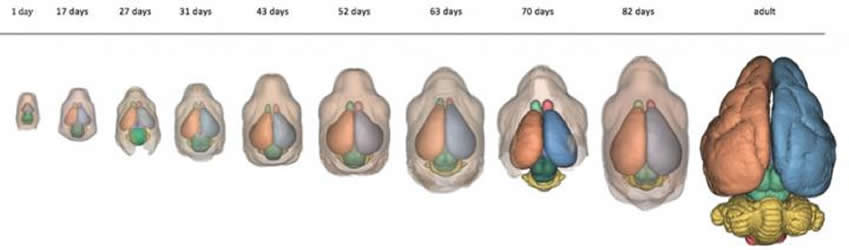
Dr Weisbecker said the research was a world first attempt at quantifying mammalian brain growth across several species.
“Instead of one neurogenesis-based rule, we suspect that the evolution of brain parts, including the huge human cerebral hemispheres, results from a complex combination of factors including the early molecular processes which divide the brain long before it starts growing,” she said.
“In addition, all parts of the brain need to be tightly interconnected, so that the broad pattern of mammalian brain partitions might be due to constraints of brain function combined with a very early, possibly species-specific, developmental brain ‘blueprint’.”
Dr Weisbecker said her lab aimed to tell the evolutionary story of today’s diverse land vertebrates through developmental biology.
“My research thrives on the huge diversity of animals that Australia has to offer,” she said.
Source: Dr. Vera Weisbecker – University of Queensland
Image Source: NeuroscienceNews.com image is credited to Dr. Vera Weisbecker.
Original Research: Full open access research for “Testing hypotheses of developmental constraints on mammalian brain partition evolution, using marsupials” by Alison Carlisle, Lynne Selwood, Lyn A. Hinds, Norman Saunders, Mark Habgood, Karine Mardon & Vera Weisbecker in Scientific Reports. Published online June 26 2017 doi:10.1038/s41598-017-02726-9
[cbtabs][cbtab title=”MLA”]University of Queensland “New Technique Challenges Brain Development Hypothesis.” NeuroscienceNews. NeuroscienceNews, 5 July 2017.
<https://neurosciencenews.com/brain-development-hypothesis-7026/>.[/cbtab][cbtab title=”APA”]University of Queensland (2017, July 5). New Technique Challenges Brain Development Hypothesis. NeuroscienceNew. Retrieved July 5, 2017 from https://neurosciencenews.com/brain-development-hypothesis-7026/[/cbtab][cbtab title=”Chicago”]University of Queensland “New Technique Challenges Brain Development Hypothesis.” https://neurosciencenews.com/brain-development-hypothesis-7026/ (accessed July 5, 2017).[/cbtab][/cbtabs]
Abstract
Testing hypotheses of developmental constraints on mammalian brain partition evolution, using marsupials
There is considerable debate about whether the partition volumes of the mammalian brain (e.g. cerebrum, cerebellum) evolve according to functional selection, or whether developmental constraints of conserved neurogenetic scheduling cause predictable partition scaling with brain size. Here we provide the first investigation of developmental constraints on partition volume growth, derived from contrast-enhanced micro-computed tomography of hydrogel-stabilized brains from three marsupial species. ANCOVAs of partition vs. brain volume scaling, as well as growth curve comparisons, do not support several hypotheses consistent with developmental constraints: brain partition growth significantly differs between species, or between developing vs. adult marsupials. Partition growth appears independent of adult brain volume, with no discernable growth spurts/lags relatable to internal structural change. Rather, adult proportion differences appear to arise through growth rate/duration heterochrony. Substantial phylogenetic signal in adult brain partitions scaling with brain volume also counters expectations of development-mediated partition scaling conservatism. However, the scaling of olfactory bulb growth is markedly irregular, consistent with suggestions that it is less constrained. The very regular partition growth curves suggest intraspecific developmental rigidity. We speculate that a rigid, possibly neuromer-model-like early molecular program might be responsible both for regular growth curves within species and impressions of a link between neurogenesis and partition evolution.
“Testing hypotheses of developmental constraints on mammalian brain partition evolution, using marsupials” by Alison Carlisle, Lynne Selwood, Lyn A. Hinds, Norman Saunders, Mark Habgood, Karine Mardon & Vera Weisbecker in Scientific Reports. Published online June 26 2017 doi:10.1038/s41598-017-02726-9