Summary: 3D images show how a baby’s brain and skull change shape during labor and delivery.
Source: PLOS
Using Magnetic Resonance Imaging (MRI), scientists have captured 3D images that show how infants’ brains and skulls change shape as they move through the birth canal just before delivery. Olivier Ami of Auvergne University in Clermont Ferrand, France, and colleagues present these findings in the open access journal PLOS ONE on May 15, 2019.
Doctors have long known that infants’ heads change shape during birth. Termed “fetal head molding,” these changes occur during the second stage of labor, when the baby leaves the uterus and moves through the birth canal. However, the details of fetal head molding remain unclear, and only one prior study has captured images of this process.
In the new study, Ami and colleagues used 3D MRI to capture detailed images of seven infants’ skulls and brains before and during the second stage of labor. The analysis revealed fetal head molding during the second stage of labor in all seven infants, with different parts of the skull overlapping to varying degrees among the infants. After birth, five of the newborns’ skull and brain shapes returned to their pre-birth state, but the changes persisted in two of the infants. Two of the three infants with the greatest degree of fetal head molding were delivered by emergency C-section, but the third was delivered vaginally with minimal expulsive efforts.
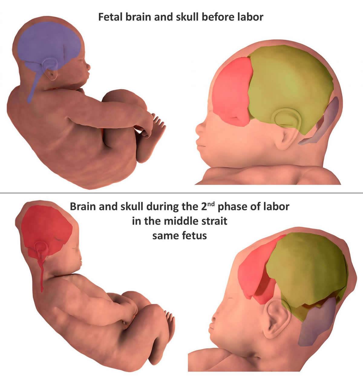
Overall, the findings suggest that infants experience greater skull stresses during birth than previously thought, potentially underlying the asymptomatic brain and retinal bleeding seen in many newborns after vaginal delivery. The authors note that a larger study is needed to confirm their findings, but that their work demonstrates the value of 3D MRI in capturing fetal head molding.
Ami adds: “During vaginal delivery, the fetal brain shape undergoes deformation to varying degrees depending on the degree of overlap of the skull bones. Fetal skull molding is no more visible in most newborns after birth. Some skulls accept the deformation (compliance) and allow an easy delivery, while others do not deform easily (non-compliance).”
Source:
PLOS
Media Contacts:
Olivier Ami – PLOS
Image Source:
The image is credited to Ami et al., 2019.
Original Research: Open access
“Three-dimensional magnetic resonance imaging of fetal head molding and brain shape changes during the second stage of labor”. Ami O, Maran J-C, Gabor P, Whitacre EB, Musset D, Dubray C, et al.
PLOS ONE. doi:10.1371/journal.pone.0215721
Abstract
Three-dimensional magnetic resonance imaging of fetal head molding and brain shape changes during the second stage of labor
To demonstrate and describe fetal head molding and brain shape changes during delivery, we used three-dimensional (3D) magnetic resonance imaging (MRI) and 3D finite element mesh reconstructions to compare the fetal head between prelabor and the second stage of labor. A total of 27 pregnant women were examined with 3D MRI sequences before going into labor using a 1 Tesla open field MRI. Seven of these patients subsequently had another set of 3D MRI sequences during the second stage of labor. Volumes of 2D images were transformed into finite element 3D reconstructions. Polygonal meshes for each part of the fetal body were used to study fetal head molding and brain shape changes. Varying degrees of fetal head molding were present in the infants of all seven patients studied during the second phase of labor compared with the images acquired before birth. The cranial deformation, however, was no longer observed after birth in five out of the seven newborns, whose post-natal cranial parameters were identical to those measured before delivery. The changing shape of the fetal brain following the molding process and constraints on the brain tissue were observed in all the fetuses. Of the three fetuses presenting the greatest molding of the skull bones and brain shape deformation, two were delivered by cesarean-section (one after a forceps failure and one for engagement default), while the fetus presenting with the greatest skull molding and brain shape deformation was born physiologically. This study demonstrates the value of 3D MRI study with 3D finite element mesh reconstruction during the second stage of labor to reveal how the fetal brain is impacted by the molding of the cranial bones. Fetal head molding was systematically observed when the fetal head was engaged between the superior pelvic strait and the middle brim.