Summary: Fear and anxiety share overlapping neural circuits. The findings contradict the popular belief that anxiety and fear are distinct emotions with different triggers and segregated brain circuits.
Source: University of Maryland
Anxiety, the most common family of mental illnesses in the U.S., has been pushed to epic new heights by the COVID-19 pandemic, with the Centers for Disease Control and Prevention estimating that nearly 1 in 3 U.S. adults and a staggering 41% of people ages 18-29 experienced clinically significant anxiety symptoms in late August. Now, the findings of a recent UMD-led study indicate that some long-accepted thinking about the basic neuroscience of anxiety is wrong.
The report by an international team of researchers led by Alexander Shackman, an associate professor of psychology at UMD, and Juyoen Hur, an assistant professor of psychology at Yonsei University in Seoul, South Korea, provides new evidence that fear and anxiety reflect overlapping brain circuits. The findings run counter to popular scientific accounts, highlighting the need for a major theoretical reckoning. The study was published last week in the Journal of Neuroscience.
“The conceptual distinction between ‘fear’ and ‘anxiety’ dates back to the time of Freud, if not the Greek philosophers of antiquity,” said Shackman, a core faculty member of UMD’s Neuroscience and Cognitive Science Program, and 2018 recipient of a seed grant award from UMD’s Brain and Behavior Initiative, “In recent years, brain imagers and clinicians have extended this distinction, arguing that fear and anxiety are orchestrated by distinct neural networks.
However, Shackman says their new study adds to a rapidly growing body of new evidence suggesting that this old mode is wrong. “If anything, fear and anxiety seem to be constructed in the brain using a massively overlapping set of neural building blocks,” he said.
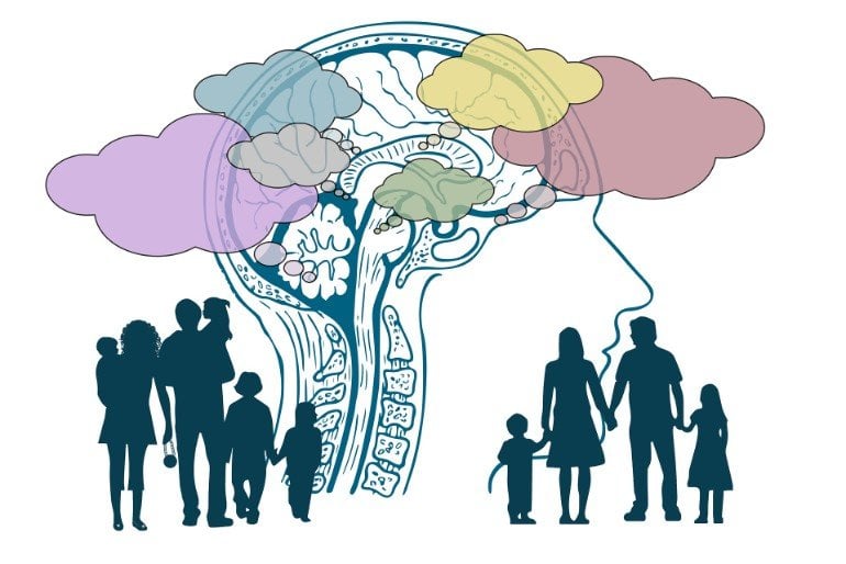
Prevailing scientific theory holds that fear and anxiety are distinct, with different triggers and strictly segregated brain circuits. Fear–a fleeting reaction to certain danger–is thought to be controlled by the amygdala, a small almond-shaped region buried beneath the wrinkled convolutions of the cerebral cortex. By contrast, anxiety–a persistent state of heightened apprehension and arousal elicited when threat is uncertain–is thought to be orchestrated by the neighboring bed nucleus of the stria terminalis (BNST). But new evidence from Shackman and his colleagues suggests that both of these brain regions are equally sensitive to certain and uncertain kinds of threats.
Leveraging cutting-edge neuroimaging techniques available at the Maryland Neuroimaging Center, their research team used fMRI to quantify neural activity while participants anticipated receiving a painful shock paired with an unpleasant image and sound–a new task that the researchers dubbed the “Maryland Threat Countdown”.
The timing of this “threat” was signaled either by a conventional countdown timer–i.e. “3, 2, 1…”–or by a random string of numbers–e.g. “16, 21, 8.” In both conditions, threat anticipation recruited a remarkably similar network of brain regions, including the amygdala and the BNST. Across a range of head-to-head comparisons, the two showed statistically indistinguishable responses.
The team examined the neural circuits engaged while waiting for certain and uncertain threat (i.e. “fear” and “anxiety”). Results demonstrated that both kinds of threat anticipation recruited a common network of core brain regions, including the amygdala and BNST.
These observations raise important questions about the Research Domain Criteria (RDoC) framework that currently guides the U.S. National Institute of Mental Health’s quest to discover the brain circuitry underlying anxiety disorders, depression, and other common mental illnesses. “As it is currently written, RDoC embodies the idea that certain and uncertain threat are processed by circuits centered on the amygdala and BNST, respectively. It’s very black-and-white thinking,” Shackman noted, emphasizing that RDoC’s “strict-segregation” model is based on data collected at the turn of the century.
“It’s time to update the RDoC so that it reflects the actual state of the science. It’s not just our study; in fact, a whole slew of mechanistic studies in rodents and monkeys, and new meta-analyses of the published human imaging literature are all coalescing around the same fundamental scientific lesson: certain and uncertain threat are processed by a shared network of brain regions, a common core,” he said.

As the crown jewel of NIMH’s strategic plan for psychiatric research in the U.S., the RDoC framework influences a wide range of biomedical stakeholders, from researchers and drug companies to private philanthropic foundations and foreign funding agencies. Shackman noted that the RDoC has an outsized impact on how fear and anxiety research is designed, interpreted, peer reviewed, and funded here in the U.S. and abroad.
“Anxiety disorders impose a substantial and growing burden on global public health and the economy,” Shackman said, “While we have made tremendous scientific progress, existing treatments are far from curative for many patients. Our hope is that research like this study can help set the stage for better models of emotion and, ultimately, hasten the development of more effective intervention strategies for the many millions of children and adults around the world who struggle with debilitating anxiety and depression.”
Funding: This work was supported by the National Institute of Mental Health and University of Maryland, College Park.
The research team also included Allegra S. Anderson, Vanderbilt University; Jinyi Kuang, University of Pennsylvania; Manuel Kuhn, Harvard Medical School; Andrew S. Fox, University of California, Davis; and Jason F. Smith, Rachael M. Tillman, Hyung Cho Kim, and Kathryn A. DeYoung, all from the University of Maryland, College Park.
About this neuroscience research news
Source: University of Maryland
Contact: Lee Tune – University of Maryland
Image: The image is in the public domain.
Original Research: Closed access.
“Anxiety and the Neurobiology of Temporally Uncertain Threat Anticipation” by Juyoen Hur, Jason F. Smith, Kathryn A. DeYoung, Allegra S. Anderson, Jinyi Kuang, Hyung Cho Kim, Rachael M. Tillman, Manuel Kuhn, Andrew S. Fox and Alexander J. Shackman. Journal of Neuroscience
Abstract
Anxiety and the Neurobiology of Temporally Uncertain Threat Anticipation
When extreme, anxiety—a state of distress and arousal prototypically evoked by uncertain danger—can be debilitating. Uncertain anticipation is a shared feature of situations that elicit signs and symptoms of anxiety across psychiatric disorders, species, and assays. Despite the profound significance of anxiety for human health and wellbeing, the neurobiology of uncertain-threat anticipation remains unsettled. Leveraging a paradigm adapted from animal research and optimized for fMRI signal decomposition, we examined the neural circuits engaged during the anticipation of temporally uncertain and certain threat in 99 men and women. Results revealed that the neural systems recruited by uncertain and certain threat anticipation are anatomically colocalized in frontocortical regions, extended amygdala, and periaqueductal gray. Comparison of the threat conditions demonstrated that this circuitry can be fractionated, with frontocortical regions showing relatively stronger engagement during the anticipation of uncertain threat, and the extended amygdala showing the reverse pattern. Although there is widespread agreement that the bed nucleus of the stria terminalis and dorsal amygdala—the two major subdivisions of the extended amygdala—play a critical role in orchestrating adaptive responses to potential danger, their precise contributions to human anxiety have remained contentious. Follow-up analyses demonstrated that these regions show statistically indistinguishable responses to temporally uncertain and certain threat anticipation. These observations provide a framework for conceptualizing anxiety and fear, for understanding the functional neuroanatomy of threat anticipation in humans, and for accelerating the development of more effective intervention strategies for pathological anxiety.
SIGNIFICANCE STATEMENT
Anxiety—an emotion prototypically associated with the anticipation of uncertain harm—has profound significance for public health, yet the underlying neurobiology remains unclear. Leveraging a novel neuroimaging paradigm in a relatively large sample, we identify a core circuit responsive to both uncertain and certain threat anticipation, and show that this circuitry can be fractionated into subdivisions with a bias for one kind of threat or the other. The extended amygdala occupies center stage in neuropsychiatric models of anxiety, but its functional architecture has remained contentious. Here we demonstrate that its major subdivisions show statistically indistinguishable responses to temporally uncertain and certain threat. Collectively, these observations indicate the need to revise how we think about the neurobiology of anxiety and fear.


