

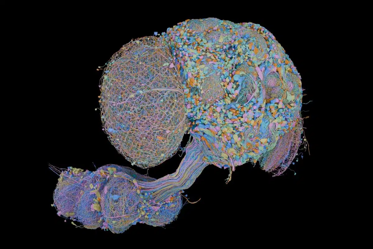
Summary:
Following nearly two decades of large-scale connectomics research, scientists have mapped every neuron and connection within the adult male fruit fly’s central nervous system, establishing a complete wiring diagram of more than 166,000 neurons. The milestone provides an unprecedented circuit-level view of how sensory cues translate into motor execution and opens the door to mapping complex vertebrate brains.
Key Facts:
- Complete CNS Circuitry: The wiring diagram reconstructs more than 166,000 neurons and millions of synaptic connections spanning the adult male fruit fly’s brain, both optic lobes, and the ventral nerve cord.
- 1,000-Fold Efficiency Gain: By developing high-resolution microscopy and AI-powered computational algorithms in collaboration with Google Research, investigators reduced the time and resources required to generate whole-brain connectomes by more than 1,000-fold.
- Stepping Stone to Vertebrate Brains: Building on this platform, researchers are now applying these methodologies to map the brains of transparent vertebrate fish—larval zebrafish and adult Danionella—to model complex behavior and neurodegenerative disorders.
Source: Howard Hughes Medical Institute (HHMI) Janelia Research Campus
Mapping 166,000 Neurons: A Two-Decade Quest
When scientists at the Howard Hughes Medical Institute’s Janelia Research Campus first announced their ambition in 2008 to chart every neuron inside the fruit fly brain, the project was met with widespread skepticism across the neuroscience community.
At the time, the only fully mapped nervous system was that of the nematode Caenorhabditis elegans, an organism with just 302 neurons whose reconstruction required more than a decade of tedious, manual labor. Charting the nervous system of an insect containing over 100,000 cells seemed prohibitively expensive, agonizingly slow, and technically out of reach.
Now, that long-term gamble has paid off. In collaboration with teams at the MRC Laboratory of Molecular Biology, the University of Cambridge, and Google Research, Janelia investigators have completed the full connectome of an adult male fruit fly (Drosophila melanogaster).
The finished blueprint maps more than 166,000 neurons and millions of synaptic intersections across the entire central nervous system, integrating the brain, both optic lobes, and the ventral nerve cord, the insect counterpart to the vertebrate spinal cord.
Credit: HHMI
“None of those things would’ve happened if we hadn’t done the fly,” said Gerry Rubin, head of Biology, senior group leader, and founding executive director at Janelia. “It was us having the leap of faith that we could assemble an interdisciplinary team who would develop ways to increase the efficiency of generating connectomes by more than 1,000-fold. That was our key contribution, and without that, we could still be waiting.”
Breaking Technical Bottlenecks with Advanced Optics and AI
When the project commenced, researchers calculated that assembling a fly connectome using standard methods would require 500 specialists working for a decade. Transforming that forecast into reality required fundamentally restructuring imaging technologies and computer vision.
Under the direction of senior group leader Harald Hess, the research group iteratively refined and scaled customized electron microscopy techniques to capture high-contrast images of individual neuronal membranes at nanoscale resolution. Simultaneously, computational teams partnered with Google Research to build machine-learning models capable of segmenting dense neural tissues and interpreting vast image datasets automatically.
This gradual scaling achieved proof-of-concept in 2020 with the release of the “hemibrain,” a detailed reconstruction of 25,000 neurons across half of the fly brain. The hemibrain demonstrated the power of connectomics, catalyzing hundreds of follow-up studies into sensory and cognitive processing across global laboratories.
From Sensory Input to Motor Output
The completion of the whole central nervous system connectome marks the first time neuroscientists can trace an unbroken, end-to-end circuit from sensory organs straight to motor effectors in an adult animal.
Credit: HHMI
Researchers can now follow pathways from the eyes, antennae, and taste receptors as signals travel into computational brain hubs and cascade downward into motor circuits within the ventral nerve cord. This continuous path allows investigators to determine the exact cellular logic that governs navigation, courtship, evasion, and predatory defense.
Furthermore, the male connectome provides a critical baseline for comparative connectomics. Researchers have already leveraged the data to explore sexually dimorphic circuits that govern mating and aggression, while delineating the fine wiring underlying insect visual processing and gustatory discrimination.
The Bridge to Vertebrate Neuroscience
The methodologies validated in Drosophila are already being directed toward vertebrate systems. Janelia researchers are actively constructing connectomes for two optically transparent vertebrate models: the larval zebrafish (Danio rerio) and the miniature adult fish Danionella cerebrum.
By deciphering the organizational architecture of these vertebrate systems at single-cell resolution, scientists aim to build mechanistic models of behavioral generation. These vertebrate wiring diagrams are expected to provide foundational frameworks for understanding human brain dysfunction, shedding light on the circuit breakdowns that drive disorders such as major depression and Alzheimer’s disease.
Editorial Notes:
- This article was edited by a Neuroscience News editor.
- Journal paper reviewed in full.
- Additional context added by our staff.
About this AI and Neuroscience Research:
- Media Contact: Halea Kerr-Layton
- Source: HHMI
- Image Credit: Image credited to FlyEM Project Team at HHMI’s Janelia Research Campus, the Cambridge Connectomics Group, and Google Research. Image by Philip Hubbard/HHMI Janelia Research Campus
- Original Research is Open Access: Cell (September 3, 2026). “Sexual dimorphism in the complete Drosophila male central nervous system connectome” Authors: Stuart Berg, Isabella R. Beckett, Marta Costa, Philipp Schlegel, Michał Januszewski, Elizabeth C. Marin, Aljoscha Nern, Stephan Preibisch, Wei Qiu, Shin-ya Takemura, Alexandra M.C. Fragniere, Andrew S. Champion, Diane-Yayra Adjavon, Michael Cook, Marina Gkantia, Kenneth J. Hayworth, Gary B. Huang, William T. Katz, Florian Kämpf, Zhiyuan Lu, Christopher Ordish, Tyler Paterson, Tomke Stürner, Eric T. Trautman, Catherine R. Whittle, Laura E. Burnett, Judith Hoeller, Feng Li, Frank Loesche, Billy J. Morris, Tobias Pietzsch, Markus W. Pleijzier, Valeria Silva, Yijie Yin, Iris Ali, Griffin Badalamente, Alexander Shakeel Bates, Rory J. Beresford, John Bogovic, Paul Brooks, Sebastian Cachero, Brandon S. Canino, Bhumpanya Chaisrisawatsuk, Jody Clements, Arthur Crowe, Inês de Haan Vicente, Georgia Dempsey, Erika Donà, Márcia Dos Santos, Marisa Dreher, Christopher R. Dunne, Katharina Eichler, Samantha Finley-May, Miriam A. Flynn, Imran Hameed, Gary Patrick Hopkins, Philip M. Hubbard, Ladann Kiassat, Julie Kovalyak, Shirley A. Lauchie, Meghan Leonard, Alanna Lohff, Kit D. Longden, Charli A. Maldonado, Ilina Moitra, Sung Soo Moon, Caroline Mooney, Eva J. Munnelly, Nneoma Okeoma, Donald J. Olbris, Anika Pai, Birava Patel, Emily M. Phillips, Stephen M. Plaza, Alana Richards, Jennifer Rivas Salinas, Ruairí J.V. Roberts, Edward M. Rogers, Ashley L. Scott, Louis A. Scuderi, Pavithraa Seenivasan, Laia Serratosa Capdevila, Claire Smith, Rob Svirskas, Satoko Takemura, Ibrahim Tastekin, Alexander Thomson, Lowell Umayam, John J. Walsh, Holly Whittome, C. Shan Xu, Emily A. Yakal, Tansy Yang, Arthur Zhao, Reed George, Viren Jain, Vivek Jayaraman, Wyatt Korff, Geoffrey W. Meissner, Sandro Romani, Jan Funke, Christopher Knecht, Stephan Saalfeld, Louis K. Scheffer, Scott Waddell, Gwyneth M. Card, Carlos Ribeiro, Michael B. Reiser, Harald F. Hess, Gerald M. Rubin, and Gregory S.X.E. Jefferis.
- DOI: 10.1016/j.cell.2026.08.015
Abstract
Sexual dimorphism in the complete Drosophila male central nervous system connectome
Sex differences in behavior exist across all animals, typically under strong genetic regulation. In Drosophila, fruitless/doublesex transcription factors identify dimorphic neurons, but their organization into functional circuits remains unclear.
We present the connectome of the entire Drosophila male central nervous system. This contains 166,700 neurons spanning the brain and nerve cord, fully proofread and annotated, including fruitless/doublesex expression and 11,710 neuron types.
We provide the first comprehensive comparison between male and female brain connectomes to synaptic resolution, finding 8,069 isomorphic, 138 dimorphic, 289 male-specific, and 71 female-specific types.
This resource enables analysis of full sensory-to-motor circuits underlying complex behaviors and the impact of dimorphic elements. Sex-specific/dimorphic neurons are concentrated in higher brain centers, while the sensory and motor periphery is largely isomorphic.
Within higher centers, male-specific connections are organized into hotspots defined by male-specific neurons or arbors. Dimorphic neurons reroute information across sexes.


