Summary: Researchers have mapped over 70,000 synaptic connections in rat neurons using a silicon chip with 4,096 microhole electrodes, significantly advancing neuronal recording technology. Unlike traditional electron microscopy, which only visualizes synapses, this method also measures connection strength, providing deeper insight into brain network function.
The chip mimics patch-clamp electrodes but at a massive scale, enabling highly sensitive intracellular recordings from thousands of neurons simultaneously. Compared to their previous nanoneedle design, this new approach captured 200 times more synaptic connections, revealing detailed characteristics of each link.
The technology could revolutionize neural mapping, offering a powerful tool for studying brain function and diseases. Researchers are now working to apply this system in live brains to further understand real-time neural communication.
Key Facts
- Massive Neural Mapping: The new silicon chip recorded 70,000+ synaptic connections from ~2,000 neurons, far surpassing previous methods.
- Improved Recording Sensitivity: Microhole electrodes enabled 90% successful intracellular recordings, vastly improving signal quality over prior designs.
- Future Live Brain Applications: Researchers aim to adapt this technology for real-time neural activity mapping in live animals.
Source: Harvard
Harvard researchers have mapped and catalogued more than 70,000 synaptic connections from about 2,000 rat neurons, using a silicon chip capable of recording small yet telltale synaptic signals from a large number of neurons.
The research, published in Nature Biomedical Engineering, is a major advance in neuronal recording and may help bring scientists a step closer to drawing a detailed synaptic connection map of the brain.
Higher-order brain functions are believed to be derived from the ways brain cells, or neurons, are connected.

Neuron-to-neuron contact points are called synapses, and scientists seek to draw synaptic connection maps that show not only which neurons connect to which other neurons, but also how strong each connection is.
While electron microscopy has been used with great success to make visual maps of synaptic connections, these images lack information on connection strengths and thus the ultimate function of the neuronal network.
In contrast, a patch-clamp electrode, the gold standard in neuronal recording, can effectively get inside an individual neuron to record a faint synaptic signal with high sensitivity, and thus can find a synaptic connection and tell its strength.
Scientists have long tried to apply such high-sensitivity intracellular recording to a large number of neurons in parallel, in order to measure and characterize a large number of synaptic signals and thus draw a map annotated with connection strengths.
But they have seldom gotten further than obtaining intracellular access from a handful of neurons at once.
The researchers, led by Donhee Ham, the John A. and Elizabeth S. Armstrong Professor of Engineering and Applied Sciences at the Harvard John A. Paulson School of Engineering and Applied Sciences (SEAS), developed an array of 4,096 microhole electrodes on a silicon chip, which performed massively parallel intracellular recording of rat neurons cultured on the chip.
From these unprecedented recording data that abounded with synaptic signals, they extracted over 70,000 synaptic connections from about 2,000 neurons.
The work builds on the team’s 2020 breakthrough device – an array of 4,096 vertical nanoneedle electrodes sticking out of a silicon chip of the same integrated circuit design.
On this previous device, a neuron could wrap around a needle to allow intracellular recording, which was parallelized through the large number of electrodes. In the best case, they could extract about 300 synaptic connections from the recording data – still blowing well past what patch-clamp recording can reach.
With the basic premise in hand, the team suspected they could do better. Co-lead authors Jun Wang and Woo-Bin Jung from the Ham group at SEAS led the design and fabrication of the microhole electrode array on the silicon chip, the electrophysiological recording and the data analysis.
They operated the chip to gently open up cells with small current injections through the electrodes in order to parallelize their intracellular recording. Postdoctoral researcher Wang said the microhole design is similar to the patch-clamp electrode, which is essentially an electrode-housing glass pipette with a hole at the end.
“Not only do microhole electrodes better couple to the interiors of neurons than the vertical nanoneedle electrodes, but they are also much easier to fabricate. This accessibility is another important feature of our work,” Wang said.
The new design exceeded the team’s expectations. On average, more than 3,600 microhole electrodes out of the total 4,096 – that is, 90 percent – were intracellularly coupled to neurons on top.
The number of synaptic connections the team extracted from such unprecedented network-wide intracellular recording data bloomed to 70,000 plausible synaptic connections, compared with about 300 with their previous nanoneedle electrode array.
The quality of the recording data was also better, which allowed the team to categorize each synaptic connection based on its characteristics and strengths.
“The integrated electronics in the silicon chip plays as equally an important role as the microhole electrode, providing gentle currents in an elaborate way to obtain intracellular access, and recording at the same time the intracellular signals,” said Jung, a former postdoctoral researcher and now a faculty member at Pohang University of Science and Technology in South Korea.
“One of the biggest challenges, after we succeeded in the massively parallel intracellular recording, was how to analyze the overwhelming amount of data,” Ham said. “We have since come a long way to gain insight into synaptic connections from them. We are now working toward a newer design that can be deployed in a live brain.”
Paper co-authors include Rona S. Gertner of the Department of Chemistry and Chemical Biology, and Hongkun Park, the Mark Hyman, Jr. Professor of Chemistry and Professor of Physics.
Funding: The research was supported by the Samsung Advanced Institute of Technology of Samsung Electronics.
About this AI and neuroscience research news
Author: Anne Manning
Source: Harvard
Contact: Anne Manning – Harvard
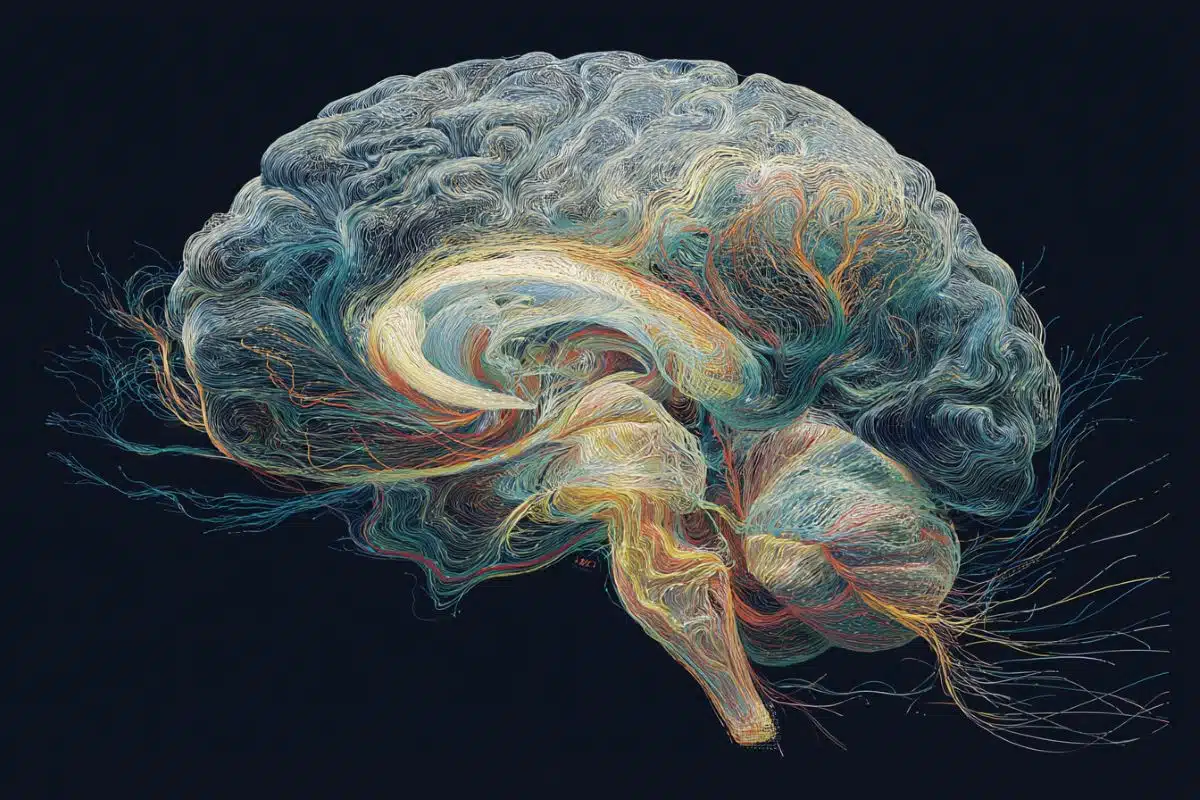
Image: The image is credited to Neuroscience News
Original Research: Closed access.
“Synaptic connectivity mapping among thousands of neurons via parallelized intracellular recording with a microhole electrode array” by Donhee Ham et al. Nature Biomedical Engineering
Abstract
Synaptic connectivity mapping among thousands of neurons via parallelized intracellular recording with a microhole electrode array
The massive parallelization of neuronal intracellular recording, which enables the measurement of synaptic signals across a neuronal network, and thus the mapping and characterization of synaptic connections, is an open challenge, with the state of the art being limited to the mapping of about 300 synaptic connections.
Here we report a 4,096 platinum/platinum-black microhole electrode array fabricated on a complementary metal-oxide semiconductor chip for parallel intracellular recording and thus for synaptic-connectivity mapping.
The microhole–neuron interface, together with current-clamp electronics in the underlying semiconductor chip, allowed a 90% average intracellular coupling rate in rat neuronal cultures, generating network-wide intracellular-recording data with abundant synaptic signals.
From these data, we extracted more than 70,000 plausible synaptic connections among more than 2,000 neurons and catalogued them into electrical synaptic connections and into inhibitory, weak/uneventful excitatory and strong/eventful excitatory chemical synaptic connections, with an estimated overall error rate of about 5%.
This scale of synaptic-connectivity mapping and the ability to characterize synaptic connections is a step towards the functional connectivity mapping of large-scale neuronal networks.