Summary: Researchers use AI to reveal distinct cellular-level differences in the brains of men and women, focusing on white matter. These findings show AI can accurately identify sex-based brain patterns invisible to human eyes.
The study suggests that understanding these differences can enhance diagnostic tools and treatments for brain disorders. This research emphasizes the need for diversity in brain studies to ensure comprehensive insights into neurological diseases.
Key Facts:
- AI Accuracy: AI models identified biological sex in MRI scans with 92%-98% accuracy.
- White Matter Focus: Differences were found in the brain’s white matter, crucial for inter-regional communication.
- Enhanced Diagnostics: Understanding sex-based brain differences can improve diagnostics and treatments for disorders like multiple sclerosis and autism.
Source: NYU Langone
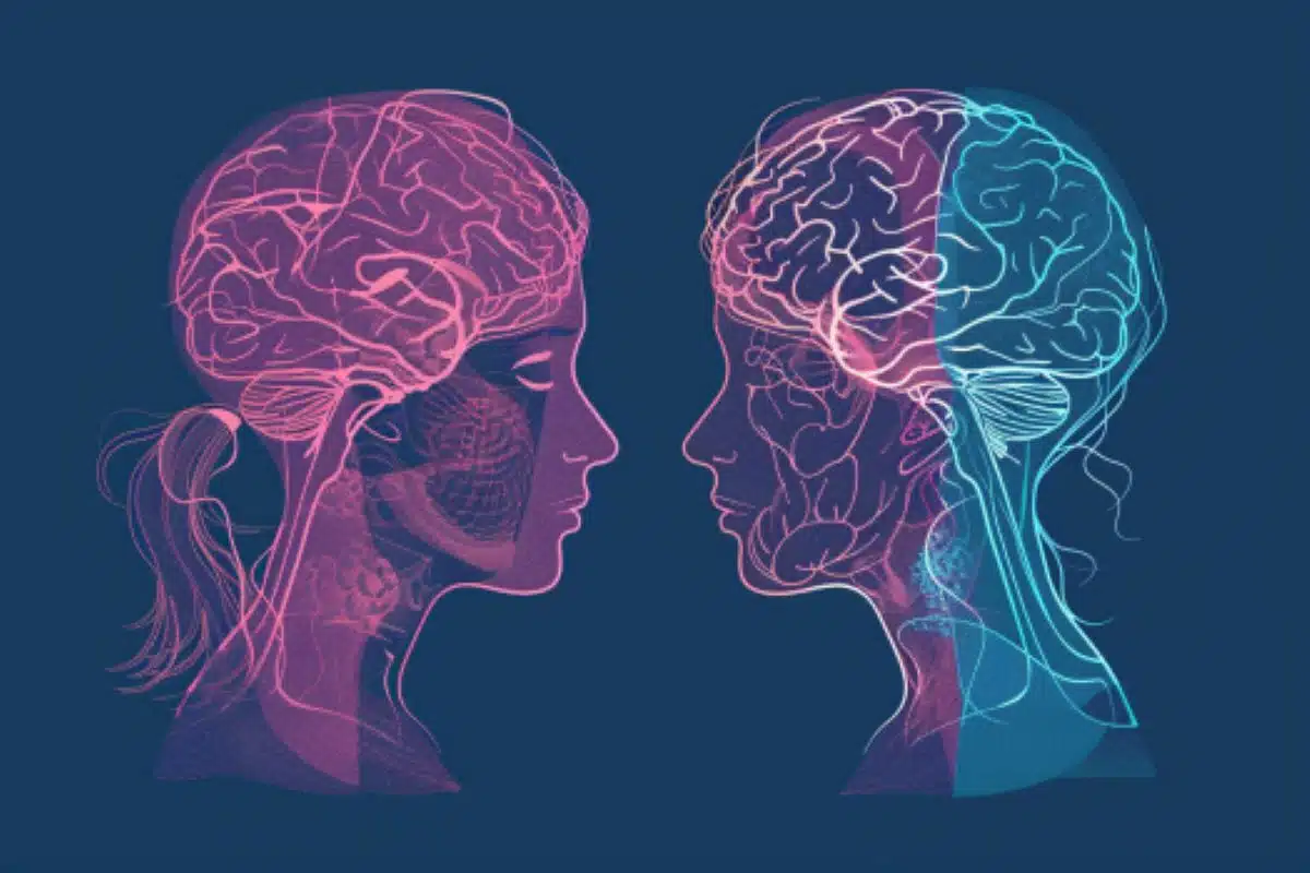
Artificial intelligence (AI) computer programs that process MRI results show differences in how the brains of men and women are organized at a cellular level, a new study shows. These variations were spotted in white matter, tissue primarily located in the human brain’s innermost layer, which fosters communication between regions.
Men and women are known to experience multiple sclerosis, autism spectrum disorder, migraines, and other brain issues at different rates and with varying symptoms.
A detailed understanding of how biological sex impacts the brain is therefore viewed as a way to improve diagnostic tools and treatments.
However, while brain size, shape, and weight have been explored, researchers have only a partial picture of the brain’s layout at the cellular level.
Led by researchers at NYU Langone Health, the new study used an AI technique called machine learning to analyze thousands of MRI brain scans from 471 men and 560 women.
Results revealed that the computer programs could accurately distinguish between biological male and female brains by spotting patterns in structure and complexity that were invisible to the human eye.
The findings were validated by three different AI models designed to identify biological sex using their relative strengths in either zeroing in on small portions of white matter or analyzing relationships across larger regions of the brain.
“Our findings provide a clearer picture of how a living, human brain is structured, which may in turn offer new insight into how many psychiatric and neurological disorders develop and why they can present differently in men and women,” said study senior author and neuroradiologist Yvonne Lui, MD.
Lui, a professor and vice chair for research in the Department of Radiology at NYU Grossman School of Medicine, notes that previous studies of brain microstructure have largely relied on animal models and human tissue samples.
In addition, the validity of some of these past findings has been called into question for relying on statistical analyses of “hand-drawn” regions of interest, meaning researchers needed to make many subjective decisions about the shape, size, and location of the regions they choose. Such choices can potentially skew the results, says Lui.
The new study results, publishing online May 14 in the journal Scientific Reports, avoided that problem by using machine learning to analyze entire groups of images without asking the computer to inspect any specific spot, which helped to remove human biases, the authors say.
For the research, the team started by feeding AI programs existing data examples of brain scans from healthy men and women and also telling the machine programs the biological sex of each brain scan.
Since these models were designed to use complex statistical and mathematical methods to get “smarter” over time as they accumulated more data, they eventually “learned” to distinguish biological sex on their own. Importantly, the programs were restricted from using overall brain size and shape to make their determinations, says Lui.
According to the results, all of the models correctly identified the sex of subject scans between 92% and 98% of the time. Several features in particular helped the machines make their determinations, including how easily and in what direction water could move through brain tissue.
“These results highlight the importance of diversity when studying diseases that arise in the human brain,” said study co-lead author Junbo Chen, MS, a doctoral candidate at NYU Tandon School of Engineering.
“If, as has been historically the case, men are used as a standard model for various disorders, researchers may miss out on critical insight,” added study co-lead author Vara Lakshmi Bayanagari, MS, a graduate research assistant at NYU Tandon School of Engineering.
Bayanagari cautions that while the AI tools could report differences in brain-cell organization, they could not reveal which sex was more likely to have which features. She adds that the study classified sex based on genetic information and only included MRIs from cis-gendered men and women.
According to the authors, the team next plans to explore the development of sex-related brain structure differences over time to better understand environmental, hormonal, and social factors that could play a role in these changes.
Funding: Funding for the study was provided by the National Institutes of Health grants R01NS119767, R01NS131458, and P41EB017183, as well as by the United States Department of Defense grant W81XWH2010699.
In addition to Lui, Chen, and Bayanagari, other NYU Langone Health and NYU researchers involved in the study were Sohae Chung, PhD, and Yao Wang, PhD.
About this AI and neuroscience research news
Author: Shira Polan
Source: NYU Langone
Contact: Shira Polan – NYU Langone
Image: The image is credited to Neuroscience News
Original Research: Open access.
“Deep Learning with Diffusion MRI as in vivo Microscope Reveals Sex-related Differences in Human White Matter Microstructure” by Yvonne Lui et al. Scientific Reports
Abstract
Deep Learning with Diffusion MRI as in vivo Microscope Reveals Sex-related Differences in Human White Matter Microstructure
Biological sex is a crucial variable in neuroscience studies where sex differences have been documented across cognitive functions and neuropsychiatric disorders.
While gross statistical differences have been previously documented in macroscopic brain structure such as cortical thickness or region size, less is understood about sex-related cellular-level microstructural differences which could provide insight into brain health and disease.
Studying these microstructural differences between men and women paves the way for understanding brain disorders and diseases that manifest differently in different sexes.
Diffusion MRI is an important in vivo, non-invasive methodology that provides a window into brain tissue microstructure.
Our study develops multiple end-to-end classification models that accurately estimates the sex of a subject using volumetric diffusion MRI data and uses these models to identify white matter regions that differ the most between men and women. 471 male and 560 female healthy subjects (age range, 22–37 years) from the Human Connectome Project are included.
Fractional anisotropy, mean diffusivity and mean kurtosis are used to capture brain tissue microstructure characteristics.
Diffusion parametric maps are registered to a standard template to reduce bias that can arise from macroscopic anatomical differences like brain size and contour.
This study employ three major model architectures: 2D convolutional neural networks, 3D convolutional neural networks and Vision Transformer (with self-supervised pertaining).
Our results show that all 3 models achieve high sex classification performance (test AUC 0.92–0.98) across all diffusion metrics indicating definitive differences in white matter tissue microstructure between males and females.
We further use complementary model architectures to inform about the pattern of detected microstructural differences and the influence of short-range versus long-range interactions.
Occlusion analysis together with Wilcoxon signed-rank test is used to determine which white matter regions contribute most to sex classification.
The results indicate that sex-related differences manifest in both local features as well as global features / longer-distance interactions of tissue microstructure.
Our highly consistent findings across models provides new insight supporting differences between male and female brain cellular-level tissue organization particularly in the central white matter.