Summary: Researchers report on the role the thalamus plays in visual processing.
Source: FMI.
Botond Roska and his team at the FMI have investigated how visual features, extracted in the eye, are combined in the thalamus, the second stage of visual processing in the brain. They have shown that the function of the thalamus is not merely to relay information. Rather, synaptic connections between different types of retinal cells and individual thalamic cells permit the integration of different visual features – in some cases from both eyes.
The sensory thalamus, a small structure located between the brain stem and the cortex, is an important gateway to the brain. Sensory information-what we see, hear, taste or feel-first passes through the thalamus before being processed in higher brain areas and the sensory thalamus was long thought to be the brain’s relay station. Recently, however, evidence has accumulated that the thalamus not only relays but also processes input. But it has not been clear what happens at the single cell level.
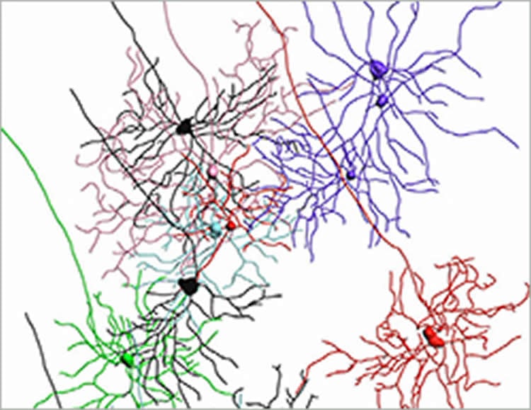
To address this question, Santiago Rompani, a postdoctoral fellow in Botond Roska’s group, developed a sophisticated method using rabies virus to track individual neuronal connections of single thalamic cells in mice. In addition, Fiona Müllner, another postdoc in Roska’s lab, developed robust statistical analysis to make sense of the data. They focused on an area known as the lateral geniculate nucleus (LGN), where input from the visual system converges. In the LGN, the axons of different types of retinal ganglion cells make synaptic connections with thalamic neurons. Botond Roska, Senior group leader at the FMI comments: “This is the first time we’ve been able to trace neuronal connections of a single cell in such a deep structure of the brain.”
Using this method, the scientists then showed that ganglion cells connect to thalamic cells in different modes. In the first mode, 1-5 ganglion cells, mostly of the same type, connect with one LGN cell. According to Roska, “This ‘relay mode’ is what we would have expected: Information from one type of ganglion cell transmitting one feature of the visual scene is further passed on.”
Interestingly, two other less expected and more prominent modes were also identified. In the second mode, 6-36 ganglion cells of different types converge from one eye; in this case, the LGN cell receives and integrates information about different aspects of the visual scene before passing it on. A function that goes beyond a pure relay function. In the third mode, a subset of LGN cells receives direct input from up to 91 ganglion cells of different types from both eyes. “This was unexpected,” says Roska. “How this integration functions and how it influences our perception remains to be elucidated.”
Roska concludes: “We now think that the thalamus takes on a much broader role in sensory information processing, and that different degrees of specialization of thalamic cells add an additional layer to how we perceive our surroundings.”
Source: FMI
Image Source: NeuroscienceNews.com image is credited to FMI.
Original Research: Full open access research “Different Modes of Visual Integration in the Lateral Geniculate Nucleus Revealed by Single-Cell-Initiated Transsynaptic Tracing” by Santiago B. Rompani, Fiona E. Müllner, Adrian Wanner, Chi Zhang, Chiara N. Roth, Keisuke Yonehara, and Botond Roska in Neuron. Published online February 23 2017 doi:10.1016/j.neuron.2017.01.028
[cbtabs][cbtab title=”MLA”]FMI “How the Thalamus Helps Us to See.” NeuroscienceNews. NeuroscienceNews, 22 February 2017.
<https://neurosciencenews.com/vision-thalamus-6152/>.[/cbtab][cbtab title=”APA”]FMI (2017, February 22). How the Thalamus Helps Us to See. NeuroscienceNew. Retrieved February 22, 2017 from https://neurosciencenews.com/vision-thalamus-6152/[/cbtab][cbtab title=”Chicago”]FMI “How the Thalamus Helps Us to See.” https://neurosciencenews.com/vision-thalamus-6152/ (accessed February 22, 2017).[/cbtab][/cbtabs]
Abstract
Different Modes of Visual Integration in the Lateral Geniculate Nucleus Revealed by Single-Cell-Initiated Transsynaptic Tracing
Highlights
•Individual LGN cells integrate retinal inputs in one of three distinct modes
•Relay-mode cells integrate inputs from few retinal ganglion cells of mostly one type
•Combination- and binocular-mode cells combine inputs from many ganglion cell types
•The three integration modes exhibit different degrees of cell-type specialization
Summary
The thalamus receives sensory input from different circuits in the periphery. How these sensory channels are integrated at the level of single thalamic cells is not well understood. We performed targeted single-cell-initiated transsynaptic tracing to label the retinal ganglion cells that provide input to individual principal cells in the mouse lateral geniculate nucleus (LGN). We identified three modes of sensory integration by single LGN cells. In the first, 1–5 ganglion cells of mostly the same type converged from one eye, indicating a relay mode. In the second, 6–36 ganglion cells of different types converged from one eye, revealing a combination mode. In the third, up to 91 ganglion cells converged from both eyes, revealing a binocular combination mode in which functionally specialized ipsilateral inputs joined broadly distributed contralateral inputs. Thus, the LGN employs at least three modes of visual input integration, each exhibiting different degrees of specialization.
“Different Modes of Visual Integration in the Lateral Geniculate Nucleus Revealed by Single-Cell-Initiated Transsynaptic Tracing” by Santiago B. Rompani, Fiona E. Müllner, Adrian Wanner, Chi Zhang, Chiara N. Roth, Keisuke Yonehara, and Botond Roska in Neuron. Published online February 23 2017 doi:10.1016/j.neuron.2017.01.028