Summary: Researchers discovered a druggable target that could prevent autonomic dysfunction after spinal cord injury. The research found that microglia cells control abnormal nerve growth and autonomic reflexes.
By depleting microglia in a mouse model, researchers prevented life-threatening complications. This finding could lead to new therapies for spinal cord injuries and other neurological conditions.
Key Facts:
- Microglia cells control abnormal nerve growth and autonomic reflexes post-injury.
- Depleting microglia in mice prevented autonomic complications after spinal cord injury.
- The discovery could lead to new treatments for dysautonomia in various neurological conditions.
Source: Ohio State University
In response to stressful or dangerous stimuli, nerve cells in the spinal cord activate involuntary, autonomic reflexes often referred to as “fight or flight” responses.
These protective responses cause changes in blood pressure and the release of stress hormones into the blood stream. Normally, these responses are short-lived and well-controlled, but this changes after a traumatic spinal cord injury.
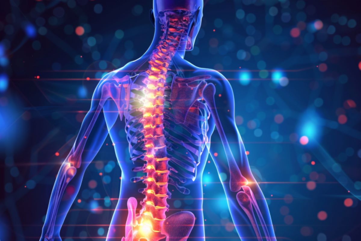
A first-ever study published in the journal Science Translational Research identifies a druggable cellular target that, if controlled properly, could prevent or lessen autonomic dysfunction and improve quality of life for people with spinal cord injury.

“We discovered that exaggerated, life-threatening autonomic reflexes after spinal cord injury are associated with abnormal growth and rewiring of nerve fibers in the spinal cord. A specific cell type, called microglia, control this abnormal growth and rewiring,” said corresponding author Phillip Popovich, PhD, professor and chair of the department of neuroscience at The Ohio State University Wexner Medical Center and College of Medicine.
“Using experimental tools to deplete microglia, we found it’s possible to prevent abnormal nerve growth, and prevent autonomic complications after spinal cord injury,” said Popovich, who also is executive director of Ohio State’s Belford Center for Spinal Cord Injury.
This research used a mouse model of spinal cord injury. However, abnormal, potentially life-threatening autonomic reflexes also occur in other animals and in people with spinal cord injury, said Popovich, who is also a member of Ohio State’s Institute for Behavioral Research Medicine.
Autonomic dysfunction or “dysautonomia,” is a major problem for people living with spinal cord injuries.
In people and animals with spinal cord injury, normally harmless stimuli, such as a full bladder, can suppress the body’s immune system and cause uncontrolled changes in blood pressure.
This leads to life threatening complications including heart attack, stroke, metabolic disease and severe infections, like pneumonia.
There is currently no treatment that prevents dysautonomia.
“We consider this a major finding,” said first author Faith Brennan, PhD, who began this work at Ohio State and is now a neuroscience researcher at Queen’s University in Kingston, Ontario. “Although this is a well-known consequence of spinal cord injury, research has mostly focused on how the injury affects neurons that control autonomic function.”
Improving autonomic function is a top priority for people living with spinal cord injury. Limiting the effects of dysautonomia after spinal cord injury would significantly increase quality of life and life expectancy, Popovich said.
Next steps for this research will focus on identifying the specific neuron-derived signals that control microglia, causing them to remodel spinal autonomic circuitry.
“Identifying these mechanisms could lead to the design of new, highly specific therapies to treat dysautonomia after spinal cord injury. It could also help in other neurological complications where dysautonomia develops, including multiple sclerosis, Alzheimer’s disease, Parkinson’s disease, stroke and traumatic brain injury,” Popovich said.
Funding: This research was supported by the National Institutes of Health, the Ray W. Poppleton Endowment, the Craig H. Neilsen Foundation and the Wings for Life Spinal Research Foundation.
About this spinal cord injury research news
Author: Eileen Scahill
Source: Ohio State University
Contact: Eileen Scahill – Ohio State University
Image: The image is credited to Neuroscience News
Original Research: Closed access.
“Microglia promote maladaptive plasticity in autonomic circuitry after spinal cord injury in mice” by Phillip Popovich et al. Science Translational Medicine
Abstract
Microglia promote maladaptive plasticity in autonomic circuitry after spinal cord injury in mice
Robust structural remodeling and synaptic plasticity occurs within spinal autonomic circuitry after severe high-level spinal cord injury (SCI). As a result, normally innocuous visceral or somatic stimuli elicit uncontrolled activation of spinal sympathetic reflexes that contribute to systemic disease and organ-specific pathology.
How hyperexcitable sympathetic circuitry forms is unknown, but local cues from neighboring glia likely help mold these maladaptive neuronal networks.
Here, we used a mouse model of SCI to show that microglia surrounded active glutamatergic interneurons and subsequently coordinated multi-segmental excitatory synaptogenesis and expansion of sympathetic networks that control immune, neuroendocrine, and cardiovascular functions.
Depleting microglia during critical periods of circuit remodeling after SCI prevented maladaptive synaptic and structural plasticity in autonomic networks, decreased the frequency and severity of autonomic dysreflexia, and prevented SCI-induced immunosuppression.
Forced turnover of microglia in microglia-depleted mice restored structural and functional indices of pathological dysautonomia, providing further evidence that microglia are key effectors of autonomic plasticity.
Additional data show that microglia-dependent autonomic plasticity required expression of triggering receptor expressed on myeloid cells 2 (Trem2) and α2δ-1–dependent synaptogenesis. These data suggest that microglia are primary effectors of autonomic neuroplasticity and dysautonomia after SCI in mice.
Manipulating microglia may be a strategy to limit autonomic complications after SCI or other forms of neurologic disease.


