

Summary: Researchers have identified how two distinct areas of the developing brain communicate and report REM sleep is key to this communication.
Source: University of Iowa.
Important interaction happens during REM sleep.
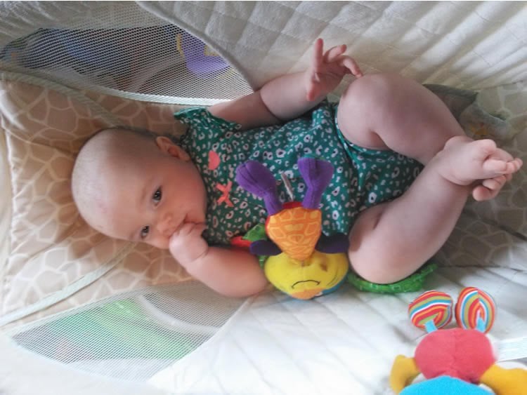
A newborn’s brain is abuzz with activity.
Day and night, it’s processing signals from all over the body, from recognizing the wriggles of the child’s own fingers and toes to the sound of mommy’s or daddy’s voice.
Though much of how the infant brain works and develops remains a mystery, University of Iowa researchers say they have uncovered a new mode of communication between two relatively distant regions. And, it turns out that sleep is key to this communication.
When two areas of the brain communicate, their rhythms will often synchronize. One well-known brain rhythm, the theta rhythm, is most closely associated with the hippocampus, a region in the forebrain important for consolidating memories and navigation, among other functions. In experiments with infant rats, the researchers showed for the first time that the hippocampus oscillates in lockstep with the red nucleus, a brain-stem structure that plays a major role in motor control. Importantly, the hippocampus and red nucleus synchronize almost exclusively during REM (active) sleep.
Rats and humans both spend much of their early lives in REM sleep. In human newborns, eight hours of every day is occupied by REM sleep alone. And because rat brains and human brains have the same basic structure, UI researchers believe the same communication, between the same regions, is likely occurring in human infants. They also suspect disruptions to that linkage may contribute to the motor-control problems that often accompany disorders such as autism and schizophrenia.
“Our findings provide a possible route to understanding the early emergence of motor problems in human infants. Because we found that communication between the hippocampus and red nucleus occurred primarily during REM sleep, disrupting normal sleep in early infancy could interfere with the strengthening of the communication links among forebrain and brainstem structures,” says Mark Blumberg, a professor in the UI Department of Psychological and Brain Sciences and corresponding author on the study, published in the journal Current Biology.
“We feel this work opens new doors to a host of important questions that have been largely overlooked,” Blumberg says.
Carlos Del Rio-Bermudez, a Fulbright Scholar who joined Blumberg’s lab for his doctoral studies, says he was surprised by the findings.
“Although a lot is known about the theta rhythm in the hippocampus and other forebrain structures, no one seems to have suspected that it might also be involved in communication between the hippocampus and a brain-stem structure like the red nucleus,” Del Rio-Bermudez says.
According to Blumberg, this discovery supports the idea that REM sleep is important for early brain development and that brain rhythms play a significant role in this process.
The researchers point out that an infant’s red nucleus and other similar structures contribute heavily to motor control at a time in development when other brain structures, including the motor cortex, are still developing.
Considering the many similarities in the brain and behavior of infant rats and humans, “it would be extraordinary if similar events are not also happening in us,” Blumberg says.
Funding: The National Institute of Child Health and Human Development and the Fulbright Foreign Student Program funded the research.
Jangjin Kim, postdoctoral research scholar, and Greta Sokoloff, research scientist in Blumberg’s lab, also contributed to the research.
Source: Richard Lewis – University of Iowa
Image Source: NeuroscienceNews.com image is credited to Neuroscience News.
Original Research: Abstract for “Theta Oscillations during Active Sleep Synchronize the Developing Rubro-Hippocampal Sensorimotor Network” by Carlos Del Rio-Bermudez, Jangjin Kim, Greta Sokoloff, and Mark S. Blumberg in Current Biology. Published online May 4 2017 doi:10.1016/j.cub.2017.03.077
[cbtabs][cbtab title=”MLA”]University of Iowa “Surprise Communication Found Between Brain Regions Involved in Infant Motor Control.” NeuroscienceNews. NeuroscienceNews, 4 May 2017.
<https://neurosciencenews.com/infant-motor-control-neurobiology-6588/>.[/cbtab][cbtab title=”APA”]University of Iowa (2017, May 4). Surprise Communication Found Between Brain Regions Involved in Infant Motor Control. NeuroscienceNew. Retrieved May 4, 2017 from https://neurosciencenews.com/infant-motor-control-neurobiology-6588/[/cbtab][cbtab title=”Chicago”]University of Iowa “Surprise Communication Found Between Brain Regions Involved in Infant Motor Control.” https://neurosciencenews.com/infant-motor-control-neurobiology-6588/ (accessed May 4, 2017).[/cbtab][/cbtabs]
Abstract
Theta Oscillations during Active Sleep Synchronize the Developing Rubro-Hippocampal Sensorimotor Network
Highlights
•Theta oscillations occur in the infant rat red nucleus (RN) during active sleep
•RN theta is first expressed as brief bursts associated with myoclonic twitching
•When tonic RN theta emerges at P12, it is tightly coupled with hippocampal theta
•Inactivation of the medial septum blocks sleep-related, but not twitch-related, theta
Summary
Neuronal oscillations comprise a fundamental mechanism by which distant neural structures establish and express functional connectivity. Long-range functional connectivity between the hippocampus and other forebrain structures is enabled by theta oscillations. Here, we show for the first time that the infant rat red nucleus (RN)—a brainstem sensorimotor structure—exhibits theta (4–7 Hz) oscillations restricted primarily to periods of active (REM) sleep. At postnatal day 8 (P8), theta is expressed as brief bursts immediately following myoclonic twitches; by P12, theta oscillations are expressed continuously across bouts of active sleep. Simultaneous recordings from the hippocampus and RN at P12 show that theta oscillations in both structures are coherent, co-modulated, and mutually interactive during active sleep. Critically, at P12, inactivation of the medial septum eliminates theta in both structures. The developmental emergence of theta-dependent functional coupling between the hippocampus and RN parallels that between the hippocampus and prefrontal cortex. Accordingly, disruptions in the early expression of theta could underlie the cognitive and sensorimotor deficits associated with neurodevelopmental disorders such as autism and schizophrenia.
“Tantrums are Not Associated with Speech or Language Deficits in Preschool Children with Autism” by Susan D. Mayes, Robin Lockridge, and Cheryl D. Tierney in Journal of Development and Physical Disabilities. Published online April 4 2017 doi:10.1007/s10882-017-9546-0


