Summary: A new study implicates structural changes in the somatosensory cortex as a cause of restless leg syndrome.
Source: AAN.
People with restless legs syndrome may have changes in a portion of the brain that processes sensory information, according to a study published in the April 25, 2018, online issue of Neurology.
Restless legs syndrome is a disorder that causes uncomfortable sensations in the legs, accompanied by an irresistible urge to move them. It often occurs in the evening and at night, sometimes affecting a person’s ability to sleep. In some cases, exercise may reduce symptoms. Iron supplements may also be prescribed if there is an iron deficiency. For more serious cases, there are also medications, but many have serious side effects if taken too long.
“Our study, which we believe is the first to show changes in the sensory system with restless legs syndrome, found evidence of structural changes in the brain’s somatosensory cortex, the area where sensations are processed,” said study author Byeong-Yeul Lee, PhD, of the University of Minnesota in Minneapolis. “It is likely that symptoms may be related to the pathological changes in this area of the brain.”
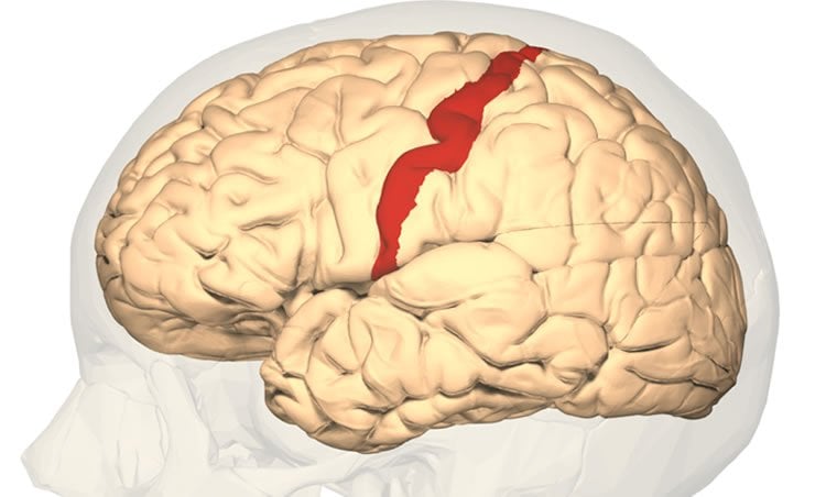
The brain’s somatosensory cortex is part of the body’s somatosensory system, which is made up of nerves and pathways that react to changes either inside or outside the body. This system helps a person perceive touch, temperature, pain, movement and position.
The study involved 28 people with severe restless legs symptoms who had the disorder for an average of 13 years. They were compared to 51 people of the same age without the disorder. Each participant had a brain scan with magnetic resonance imaging (MRI).
Researchers found that people with restless legs syndrome had a 7.5 percent decrease in the average thickness of brain tissue in the area of the brain that processes sensations compared to the healthy participants. They also found a substantial decrease in the area of the brain where nerve fibers connect one side of the brain to the other.

Lee said, “These structural changes make it even more convincing that RLS symptoms are stemming from unique changes in the brain and provide a new area of focus to understand the syndrome and possibly develop new therapies.”
He said while the study shows a possible link between symptoms and the areas of the brain that process sensory information, it is possible that symptoms may instead be linked to impaired function in other parts of the sensory system.
Funding: The study was supported by the Pennsylvania Department of Health.
Source: AAN
Publisher: Organized by NeuroscienceNews.com.
Image Source: NeuroscienceNews.com image is credited to Database Center for Life Science. Licensed CC-BY-SA-2.1-jp.
Original Research: Abstract for “Involvement of the central somatosensory system in restless legs syndrome: A neuroimaging study” by Byeong-Yeul Lee, Jongmyeong Kim, James R. Connor, Gerald D. Podskalny, Yeunchul Ryu and Qing X. Yang in Neurology. Published April 24 2018.
doi:10.1212/WNL.0000000000005562
[cbtabs][cbtab title=”MLA”]AAN “Brain Structure Linked to Symptoms of Restless Leg Syndrome.” NeuroscienceNews. NeuroscienceNews, 28 April 2018.
<https://neurosciencenews.com/somatosensory-cortex-restless-leg-8908/>.[/cbtab][cbtab title=”APA”]AAN (2018, April 28). Brain Structure Linked to Symptoms of Restless Leg Syndrome. NeuroscienceNews. Retrieved April 28, 2018 from https://neurosciencenews.com/somatosensory-cortex-restless-leg-8908/[/cbtab][cbtab title=”Chicago”]AAN “Brain Structure Linked to Symptoms of Restless Leg Syndrome.” https://neurosciencenews.com/somatosensory-cortex-restless-leg-8908/ (accessed April 28, 2018).[/cbtab][/cbtabs]
Abstract
Involvement of the central somatosensory system in restless legs syndrome: A neuroimaging study
Objective To investigate morphologic changes in the somatosensory cortex and the thickness of the corpus callosum subdivisions that provide interhemispheric connections between the 2 somatosensory cortical areas.
Methods Twenty-eight patients with severe restless legs syndrome (RLS) symptoms and 51 age-matched healthy controls were examined with high-resolution MRI at 3.0 tesla. The vertex-wise analysis in conjunction with a novel cortical surface classification method was performed to assess the cortical thickness across the whole-brain structures. In addition, the thickness of the midbody of the corpus callosum that links postcentral gyri in the 2 hemispheres was measured.
Results We demonstrated that a morphologic change occurred in the brain somatosensory system in patients with RLS compared to controls. Patients with RLS exhibited a 7.5% decrease in average cortical thickness in the bilateral postcentral gyrus (p < 0.0001). Accordingly, there was a substantial decrease in the corpus callosum posterior midbody (p < 0.008) wherein the callosal fibers are connected to the postcentral gyrus, suggesting altered white matter properties in the somatosensory pathway.
Conclusion Our results provide in vivo evidence of morphologic changes in the primary somatosensory system, which could be responsible for the sensory functional symptoms of RLS. These results provide a better understanding of the pathophysiology underlying the RLS sensory symptoms and could lead to a potential imaging marker for RLS.


