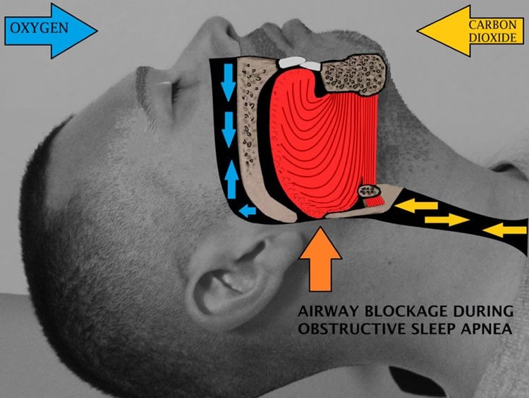
UCLA scientists develop a 3-D model of a human airway.
Imagine that before performing surgery, doctors could consult software that would determine the actual effectiveness of the procedure before even lifting a scalpel.
With the use of a computational model of the human airway being developed by Jeff Eldredge, a professor at the Henry Samueli School of Engineering and Applied Sciences at UCLA, people who suffer from sleep apnea may one day benefit from such a scenario.
Previously, Eldredge, a professor of mechanical and aerospace engineering, had been working on creating models that simulated the interactions between blood and vessel walls with Shao-Ching Huang, an expert in high performance computing from the UCLA Institute for Digital Research and Education (IDRE), which funded development of the computational tools for the project.
Last December at a conference in fluid dynamics, Eldredge and his UCLA colleagues presented the first detailed simulation of a human leg being injured by flying shrapnel, gushing blood and all. For that project, Eldredge worked in collaboration with a team from the Center for Advanced Surgical and Interventional Technology at the David Geffen School of Medicine. The goal is to train combat medics on a virtual patient that reacts in realistic ways. Researchers took CT scans of a patient’s leg to create the simulated one.
Eldredge’s current work focuses on the problem of sleep apnea — the repetitive or partial obstruction of the airway during sleep that can lead to problems including high blood pressure, stroke, and heart failure.
The UCLA School of Dentistry initially approached Eldredge about finding a way to test whether or not mandibular devices — devices that open the airway by moving the lower jaw forward — would work without having to do expensive testing where patients would have to be sent home to test out the product for weeks at a time.
Working closely with Dr. Sanjay Mallya, associate professor of dentistry, and then-UCLA medical resident Dr. Susan White, Eldredge and his partners developed a tool that simulates air-tissue interactions in the upper airway of patients. An essential piece of the tool, a high performance air flow simulation code, was developed primarily at IDRE by Huang.
The tool functions by mapping the geometry of the patient’s airway through the use of dental cone-beam CT scans. Researchers can then simulate airflow inside 3-D models of the rigid upper airways of patients with obstructive sleep apnea. By altering the geometry of the model, such as shaving a little bit off a jaw bone in the simulation, Eldredge said the computation can show what effect surgery could have on a patient without actually operating.
“You don’t want to ever have to rely on actually going in and cutting a patient to see if it gives you answers or not,” said the engineer.

While he has only been working on the issue for approximately three years, he said researchers have attempted to create such models over the last 10 to 15 years. Although these studies utilized complex geometry and real patient data, the problem has been that many of these efforts have imagined the airway as rigid — basically a pipe where air comes in as you are breathing — rather than a tissue that experiences elastic deformation and collapse as in patients with sleep apnea.
“We decided ‘Let’s build a computational tool that will actually account for the tissue collapsing,’ the actual motion of the tissue,” he said. “That meant we had to simulate the elastic deformation in the tissue of the airway and also the fluid dynamics of the air going through the airway.”
Eldredge said he views the project as a series of “baby steps.” One major goal of the research is to be able to use the computational analysis gained from his tools as a predictive tool for sleep apnea.
On the horizon, scientists may one day be able to use these tools to create a comprehensive model of the lungs as well as the upper airway.
Source: Nico Viele – UCLA
Image Source: The image is credited to Drcamachoent and is licensed CC BY SA 4.0.


