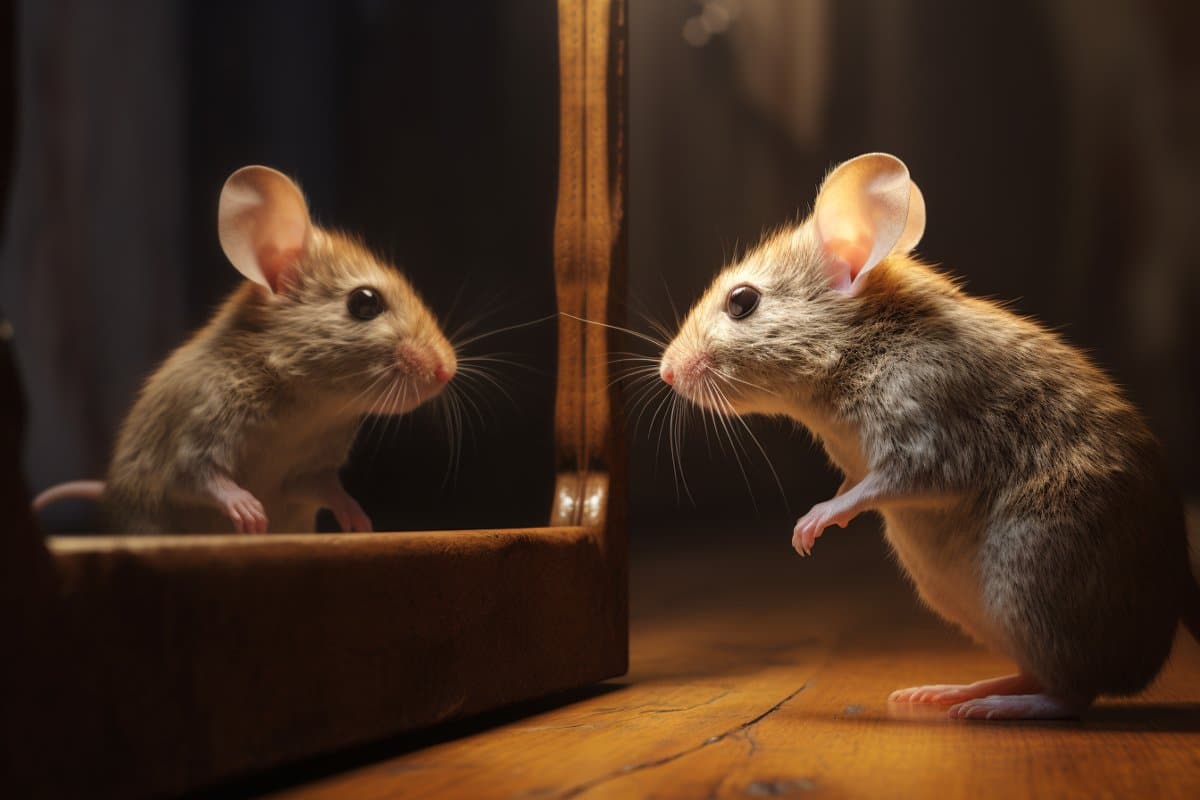
Summary: Mice display behavior akin to self-recognition when viewing their reflections in mirrors. This behavior emerges under specific conditions: familiarity with mirrors, socialization with similar-looking mice, and visible markings on their fur.
The study also identifies a subset of neurons in the hippocampus that are crucial for this self-recognition-like behavior. These findings provide valuable insights into the neural mechanisms behind self-recognition, a previously enigmatic aspect of neurobehavioral research.
Key Facts:
- Conditional Self-Recognition: Mice exhibited increased grooming behavior in response to visible white ink spots on their fur while viewing mirrors, but only when familiar with mirrors and socialized with similar-looking mice.
- Neural Mechanisms Identified: A specific group of neurons in the ventral hippocampus was found to be integral for this mirror-induced self-recognition-like behavior.
- Social and Sensory Influences: The study highlights the importance of social experiences and sensory cues in developing self-recognition capabilities, expanding our understanding of how these factors contribute to neural development.
Source: Cell Press
Researchers report December 5 in the journal Neuron that mice display behavior that resembles self-recognition when they see themselves in the mirror. When the researchers marked the foreheads of black-furred mice with a spot of white ink, the mice spent more time grooming their heads in front of the mirror—presumably to try and wash away the ink spot.
However, the mice only showed this self-recognition-like behavior if they were already accustomed to mirrors, if they had socialized with other mice who looked like them, and if the ink spot was relatively large.

The team identified a subset of neurons in the hippocampus that are involved in developing and storing this visual self-image, providing a first glimpse of the neural mechanisms behind self-recognition, something that was previously a black box in neurobehavioral research.
“To form episodic memory, for example, of events in our daily life, brains form and store information about where, what, when, and who, and the most important component is self-information or status,” says neuroscientist and senior author Takashi Kitamura of University of Texas Southwestern Medical Center.
“Researchers usually examine how the brain encodes or recognizes others, but the self-information aspect is unclear.”
The researchers used a mirror test to investigate whether mice could detect a change in their own appearance—in this case, a dollop of ink on their foreheads. Because the ink also provided a tactile stimulus, the researchers tested the black-furred mice with both black and white ink.
Though the mirror test was originally developed to test consciousness in different species, the authors note that their experiments only show that mice can detect a change in their own appearance, but this does not necessarily mean that they are “self-aware.”
They found that mice could indeed detect changes to their appearance, but only under certain conditions. Mice who were familiar with mirrors spent significantly more time grooming their heads (but not other parts of their bodies) in front of the mirror when they were marked with dollops of white ink that were 0.6 cm2 or 2 cm2.
However, the mice did not engage in increased head grooming when the ink was black—the same color as their fur—or when the ink mark was small (0.2 cm2), even if the ink was white, and mice who were not habituated to mirrors before the ink test did not display increased head grooming in any scenario.
“The mice required significant external sensory cues to pass the mirror test—we have to put a lot of ink on their heads, and then the tactile stimulus coming from the ink somehow enables the animal to detect the ink on their heads via a mirror reflection,” says first author Jun Yokose of University of Texas Southwestern Medical Center. “Chimps and humans don’t need any of that extra sensory stimulus.”
Using gene expression mapping, the researchers identified a subset of neurons in the ventral hippocampus that were activated when the mice “recognized” themselves in the mirror. When the researchers selectively rendered these neurons non-functional, the mice no longer displayed the mirror-and-ink-induced grooming behavior.
A subset of these self-responding neurons also became activated when the mice observed other mice of the same strain (and therefore similar physical appearance and fur color), but not when they observed a different strain of mouse that had white fur.
Because previous studies in chimpanzees have suggested that social experience is required for mirror self-recognition, the researchers also tested mice who had been socially isolated after weaning. These socially isolated mice did not display increased head grooming behavior during the ink test, and neither did black-furred mice that were reared alongside white-furred mice.
The gene expression analysis also showed that socially isolated mice did not develop self-responding neuron activity in the hippocampus, and neither did the black-furred mice that were reared by white-furred mice, suggesting that mice need to have social experiences alongside other similar-looking mice in order to develop the neural circuits required for self-recognition.
“A subset of these self-responding neurons was also reactivated when we exposed the mice to other individuals of the same strain,” says Kitamura.
“This is consistent with previous human literature that showed that some hippocampal cells fire not only when the person is looking at themselves, but also when they look at familiar people like a parent.”
Next, the researchers plan to try to disentangle the importance of visual and tactile stimuli to test whether mice can recognize changes in their reflection in the absence of a tactile stimulus—perhaps by using technology similar to the filters on social media apps that allow people to give themselves puppy-dog faces or bunny ears.
They also plan to study other brain regions that might be involved in self-recognition and to investigate how the different regions communicate and integrate information.
“Now that we have this mouse model, we can manipulate or monitor neural activity to comprehensively investigate the neural circuit mechanisms behind how self-recognition-like behavior is induced in mice,” says Yokose.
Funding: This research was supported by the Endowed Scholar Program, the Brain & Behavior Research Foundation, the Daiichi Sankyo Foundation of Life Science, and Uehara Memorial Foundation.
About this neuroscience research news
Author: Kristopher Benke
Source: Cell Press
Contact: Kristopher Benke – Cell Press
Image: The image is credited to Neuroscience News
Original Research: Open access.
“Visuotactile integration facilitates mirror-induced self-directed behavior through activation of hippocampal neuronal ensembles in mice” by Takashi Kitamura et al. Neuron
Abstract
Visuotactile integration facilitates mirror-induced self-directed behavior through activation of hippocampal neuronal ensembles in mice
Highlights
- Visuotactile stimuli facilitate mirror-induced self-directed behavior (MSB) in mice
- Social experience with a same-strain conspecific and mirror habituation facilitate MSB
- A subset of ventral hippocampal CA1 (vCA1) neurons responds to self and elicits MSB
- Self-responding vCA1 neurons respond to same, but not different, strain conspecifics
Summary
Remembering the visual features of oneself is critical for self-recognition. However, the neural mechanisms of how the visual self-image is developed remain unknown because of the limited availability of behavioral paradigms in experimental animals.
Here, we demonstrate a mirror-induced self-directed behavior (MSB) in mice, resembling visual self-recognition. Mice displayed increased mark-directed grooming to remove ink placed on their heads when an ink-induced visual-tactile stimulus contingency occurred. MSB required mirror habituation and social experience.
The chemogenetic inhibition of dorsal or ventral hippocampal CA1 (vCA1) neurons attenuated MSB. Especially, a subset of vCA1 neurons activated during the mirror exposure was significantly reactivated during re-exposure to the mirror and was necessary for MSB.
The self-responding vCA1 neurons were also reactivated when mice were exposed to a conspecific of the same strain.
These results suggest that visual self-image may be developed through social experience and mirror habituation and stored in a subset of vCA1 neurons.


