Summary: A new study in fruit fly models reveals STRIPAK components act as a switch to turn off quiescence and turn on the reactivation of neural stem cells.
Source: University of Plymouth
Our brains are notoriously bad at regenerating cells that have been lost through injury or disease. While therapies using neural stem cells (NSCs) hold the promise of replacing lost cells, scientists need to better understand how NSCs behave in the brain in order to develop effective treatments.
Now research led by the University of Plymouth helps to shed new light on the mechanisms used by NSCs to ‘wake up’ – going from their usual dormant state to one of action.
NSCs produce neurons (nerve cells) and surrounding glial cells in the brain. By understanding how NSCs work, it could pave the way for therapies to speed up the neurons’ and glial cells’ regeneration.
The new study, conducted using Drosophila fruit flies, shows that molecules that form a complex called STRIPAK are essential to promote reactivation in NSCs. STRIPAK (Striatin-interacting phosphatase and kinase) is found in organisms from fungi to humans, and the team uncovered it when comparing the genetic messages of dormant and reactivated NSCs in live fly brains.
The researchers then discovered that STRIPAK components act as a switch to turn off dormancy (or quiescence) and turn on reactivation.
Lead author Dr Claudia Barros, from the Institute of Translational and Stratified Medicine at the University of Plymouth, acknowledges there is still a long way to go until such findings can be translated into human treatments. But she explains the significance of the new work:
“So little is currently known about how neural stem cells coordinate cues to become active and direct the production of more brain cells,” she said. “These stem cells last throughout life mainly in a dormant state, so learning how they work is critical to our understanding of cell regeneration.

“This study reveals that STRIPAK molecules are essential to enable reactivation in NSCs, and we are very pleased with the outcomes. But we are only at the beginning. We are working to expand our findings and bring us closer to the day when human neural stem cells can be controlled and efficiently used to facilitate brain damage repair, or even prevent brain cancer growth that is fuelled by stem-like cells.”
Funding: The work was supported by the University of Plymouth, Faculty of Medicine and Dentistry; the Leverhulme Trust; the Biotechnology and Biological Sciences Research Council (BBSRC) the DFG German Research Foundation and the Johannes Gutenberg University, Germany.
The work of Dr Barros and her team takes place within the Brain Tumour Research Centre of Excellence at the University of Plymouth.
Source:
University of Plymouth
Media Contacts:
Amy King – University of Plymouth
Image Source:
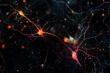
The image is credited to Dr. Claudia Barros, University of Plymouth.
Original Research: Open access
“STRIPAK Members Orchestrate Hippo and Insulin Receptor Signaling to Promote Neural Stem Cell Reactivation”. Jon Gil-Ranedo, Eleanor Gonzaga, Karolina J. Jaworek, Christian Berger, Torsten Bossing, Claudia S. Barros.
Cell Reports. doi:10.1016/j.celrep.2019.05.023
Abstract
STRIPAK Members Orchestrate Hippo and Insulin Receptor Signaling to Promote Neural Stem Cell Reactivationa
Highlights
• Transcriptional profiling of reactivating versus quiescent NSCs identifies STRIPAK members
• PP2A/Mts phosphatase inhibits Akt activation, maintaining NSC quiescence
• Mob4 and Cka target Mts to Hippo to inhibit its activity and promote NSC reactivation
• Mob4/Cka/Mts coordinate Hippo and InR/PI3K/Akt signaling in NSCs
Summary
Adult stem cells reactivate from quiescence to maintain tissue homeostasis and in response to injury. How the underlying regulatory signals are integrated is largely unknown. Drosophila neural stem cells (NSCs) also leave quiescence to generate adult neurons and glia, a process that is dependent on Hippo signaling inhibition and activation of the insulin-like receptor (InR)/PI3K/Akt cascade. We performed a transcriptome analysis of individual quiescent and reactivating NSCs harvested directly from Drosophila brains and identified the conserved STRIPAK complex members mob4, cka, and PP2A (microtubule star, mts). We show that PP2A/Mts phosphatase, with its regulatory subunit Widerborst, maintains NSC quiescence, preventing premature activation of InR/PI3K/Akt signaling. Conversely, an increase in Mob4 and Cka levels promotes NSC reactivation. Mob4 and Cka are essential to recruit PP2A/Mts into a complex with Hippo kinase, resulting in Hippo pathway inhibition. We propose that Mob4/Cka/Mts functions as an intrinsic molecular switch coordinating Hippo and InR/PI3K/Akt pathways and enabling NSC reactivation.


