

Summary: Combining advance microscopy techniques and artificial intelligence, researchers reconstruct the entire vascular network of a mouse brain down to its finest details.
Source: Helmholtz Zentrum München
Diseases of the brain are often associated with typical vascular changes. Now, scientists at Helmholtz Zentrum München, LMU University Hospital Munich and the Technical University of Munich have come up with a technique for visualising the structures of all the brain’s blood vessels – right down to the finest capillaries – including any pathological changes. So far, they have used the technique, which is based on a combination of biochemical methods and artificial intelligence, to capture the whole brain vasculature of a mouse.
Changes in the blood vessels are a hallmark of numerous brain disorders – from traumatic brain injury to stroke. Even diseases such as Alzheimer’s show changes in the fine capillaries. In short, analysing the blood vessels is key to understanding both, normal and pathological brain function. “Now we have come much closer to achieving that goal”, explains Ali Ertürk, Director of the Institute for Tissue Engineering and Regenerative Medicine at Helmholtz Zentrum München and Principal Investigator at the Institute for Stroke and Dementia Research at the LMU University Hospital Munich.
Making organs transparent
As a first step, Ertürk’s team succeeded in visualising the vascular system of mouse brains with high-resolution fluorescent microscopy without having to cut up the specimens into small sections. In order to do this, they refined the technique of tissue cleaning, in which biological tissues are treated with special dyes to render them transparent for fluorescent microscopy. “Previously, this technique could only be used to scan either the large vessels of the brain or the small ones”, says Mihail Ivilinov Todorov, a doctoral student studying under Ertürk.
Therefore the Munich-based scientists took the new approach of combining two dyes. “That gave us some great images of the brain vasculature including the capillaries”, adds the biologist.
Vascular network captured using artificial intelligence
Applying artificial intelligence, researchers from the team led by Björn Menze, Professor for Machine Learning in Biomedical Imaging at the Technical University of Munich, used these images to reconstruct the entire vascular network of the brain right down to its finest details. Such a reconstruction yields more than just images – it also allows a quantitative analysis of the vascular structures. “For example, we can statistically record the diameters of the various blood vessels or their bifurcations for different areas of the brain”, says Johannes Paetzold, doctoral student in Menze’s group.
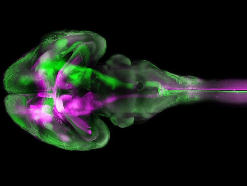
“Over the past few years, we have developed a deep learning algorithm that specialises in detecting blood vessels in medical images”, Menze explains. “This was the first time we applied it to a whole brain.” The algorithm was able to reliably distinguish between blood vessels and other tissue even though some areas in the original fluorescence images were not well-illuminated and some details were distorted due to light reflections or other errors.
Understanding and diagnosing brain disorders
Mihail Ivilinov Todorov plans to use the statistical data in order to investigate vascular changes caused by stroke, while Björn Menze is looking to study the global structures of the vascular system in order to understand the role of anatomical differences in brain dis-orders, for example.
Benefits for the patient
The method could also be used in everyday clinical practice: “With our system, we are likely to be able to analyse the small tissue specimens from human tumours with greater accuracy”, Ertürk asserts. Cancerous tissue is permeated by blood vessels, and analysing their structure helps in staging a tumour. “This may have an optimising effect on treatment”, Ertürk adds. The biologist also plans to use the new method to realise his vision for the future: the production of human organs on a 3D printer. For that to happen, a knowledge of the organ’s precise vascular structure – among many other things – will be vital.
NeuroscienceNews would like to thank Marin Bralo for submitting this article for inclusion on the website.
Source:
Helmholtz Zentrum München
Media Contacts:
Marin Bralo – Helmholtz Zentrum München
Image Source:
The image is credited to Ertürk Lab/ISD.
Original Research: Closed access
“Automa-ted analysis of whole brain vasculature using machine learning”. Mihail Ivilinov Todorov, Johannes C. Paetzold et al.
Nature Methods doi:10.1038/s41592-020-0792-1
Abstract
Automa-ted analysis of whole brain vasculature using machine learning
Tissue clearing methods enable the imaging of biological specimens without sectioning. However, reliable and scalable analysis of large imaging datasets in three dimensions remains a challenge. Here we developed a deep learning-based framework to quantify and analyze brain vasculature, named Vessel Segmentation & Analysis Pipeline (VesSAP). Our pipeline uses a convolutional neural network (CNN) with a transfer learning approach for segmentation and achieves human-level accuracy. By using VesSAP, we analyzed the vascular features of whole C57BL/6J, CD1 and BALB/c mouse brains at the micrometer scale after registering them to the Allen mouse brain atlas. We report evidence of secondary intracranial collateral vascularization in CD1 mice and find reduced vascularization of the brainstem in comparison to the cerebrum. Thus, VesSAP enables unbiased and scalable quantifications of the angioarchitecture of cleared mouse brains and yields biological insights into the vascular function of the brain.


