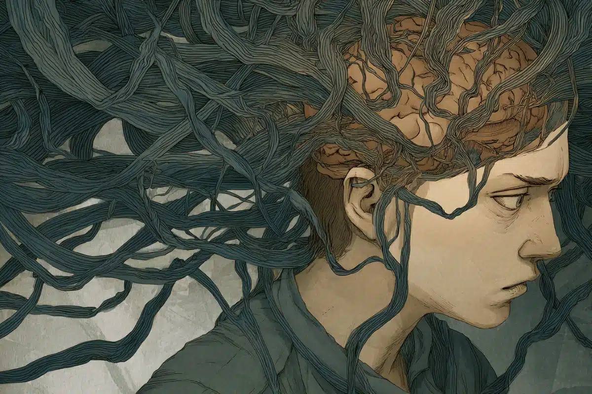
Summary: A new study reveals how specific brain cells in the hippocampus and entorhinal cortex encode time and experiences, helping us form memories and predict future outcomes.
These neurons fire in patterns reflecting the order of events, even replaying them after the experience is over. This discovery provides insight into how the brain integrates “what” and “when” information to form lasting memories.
Key Facts:
- Neurons in the hippocampus and entorhinal cortex store time-related patterns.
- The brain replays event sequences during rest, aiding memory formation.
- This finding can inform memory-enhancing neuro-prosthetic devices.
Source: UCLA
A landmark study led by UCLA Health has begun to unravel one of the fundamental mysteries in neuroscience – how the human brain encodes and makes sense of the flow of time and experiences.
The study, published in the journal Nature, directly recorded the activity of individual neurons in humans and found specific types of brain cells fired in a way that mostly mirrored the order and structure of a person’s experience.
They found the brain retains these unique firing patterns after the experience is concluded and can rapidly replay them while at rest. Furthermore, the brain is also able to utilize these learned patterns to ready itself for future stimuli following that experience.
These findings provide the first empirical evidence regarding how specific brain cells integrate “what” and “when” information to extract and retain representations of experiences through time.
The study’s senior author, Dr. Itzhak Fried, said the results could serve in the development of neuro-prosthetic devices to enhance memory and other cognitive functions as well as have implications in artificial intelligence’s understanding of cognition in the human brain.
“Recognizing patterns from experiences over time is crucial for the human brain to form memory, predict potential future outcomes and guide behaviors,” said Fried, director of epilepsy surgery at UCLA Health and professor of neurosurgery, psychiatry and biobehavioral sciences at the David Geffen School of Medicine at UCLA.
“But how this process is carried out in the brain at the cellular level had remained unknown – until now.”
Previous research, including by Dr. Fried, used brain recordings and neuroimaging to understand how the brain processes spatial navigation, showing in animal and human models that two regions of the brain – the hippocampus and the entorhinal cortex – played key roles. The two brain regions, both important in memory functions, work to interact to create a “cognitive map.”
The hippocampal neurons act as “place cells” that show when an animal is at a specific location, similar to an ‘X’ on a map, while the entorhinal neurons act as “grid cells” to provide a metric of spatial distance. These cells found first in rodents were later found in humans by Fried’s group.
Further studies have found similar neural actions work to represent non-spatial experiences such as time, sound frequency and characteristics of objects. A seminal finding by Fried and his colleagues was that of “concept cells” in human hippocampus and entorhinal cortex that responds to particular individuals, places or distinct objects and appear to be fundamental to our ability for memory.
To examine the brain processing of events in time, the UCLA study recruited 17 participants with intractable epilepsy who had been previously had depth electrodes implanted in their brains for clinical treatment.
Researchers recorded the neural activity of the participants as they underwent a complex procedure that involved behavioral tasks, pattern recognition and image sequencing.
Participants first underwent an initial screening section during which approximately 120 images of people, animals, objects and landmarks were repeatedly shown to them on a computer over about 40 minutes.
The participants were instructed to perform various tasks such as determining whether the image showed a person or not. The images, of things like famous actors, musicians and places, were selected partly based on each participant’s preferences.
Following this, the participants underwent a three-phase experiment in which they would perform behavioral tasks in response to images that were arbitrarily displayed on different locations of a pyramid-shaped graph. Six images were selected for each participant.
In the first phase, images were displayed in a pseudo-random order. The next phase had the order of images determined by the location on the pyramid graph. The final phase was identical to the first phase.
While watching these images, the participants were asked to perform various behavioral tasks that were unrelated to the positioning of the images on the pyramid graph.
These tasks included determining whether the image showed a male or female or whether a given image was mirrored compared to the previous phase.
In their analyses, Fried and his colleagues found the hippocampal-entorhinal neurons gradually began to modify and closely align their activity to the sequencing of images on the pyramid graphs.
These patterns were formed naturally and without direct instruction to the participants, according to Fried. Additionally, the neuronal patterns reflected the probability of upcoming stimuli and retained the encoded patterns even after the task was completed.
Lead author of the study was Pawel Tacikowski with co-authors Guldamla Kalendar and Davide Ciliberti.
“This study shows us for the first time how the brain uses analogous mechanisms to represent what are seemingly very different types of information: space and time,” Fried said. “We have demonstrated at the neuronal level how these representations of object trajectories in time are incorporated by the human hippocampal-entorhinal system.”
About this neuroscience and time perception research news
Author: Will Houston
Source: UCLA
Contact: Will Houston – UCLA
Image: The image is credited to Neuroscience News
Original Research: The findings will appear in Nature