Summary: A new blood test that uses atomic force microscopy technology appears to be effective as an Alzheimer’s diagnostic test.
Source: EMPA
If the suspicion of Alzheimer’s disease creeps up, those affected must prepare themselves for lengthy and complex procedures until the case is clear.
A team from Empa and the Cantonal Hospital of St. Gallen is now in the process of developing a blood test that will enable a reliable diagnosis using atomic force microscopy (AFM). The researchers have recently published their first results of a successful pilot study in the journal Science Advances.
A deep look into the molecular universe
In the beginning, physicist Peter Nirmalraj wanted to understand the molecular pathogenesis of Alzheimer’s in order to enable new approaches in diagnostics and therapy.
One step further would be to decipher the exact role of beta-amyloid peptides and tau proteins associated with the neurodegenerative disease. Nirmalraj therefore set out not only to detect the mere presence of the suspicious proteins, but also to determine their variable shape and form as well as their amounts.
Current methods allow the determination of the total amount of both proteins in body fluids. However, these techniques do not allow the visualization of differences in the shape and condition of protein accumulations. The researcher is therefore working on technologies that allow nanometer-scale observations in blood and yet do not destroy the structure and morphology of the proteins.
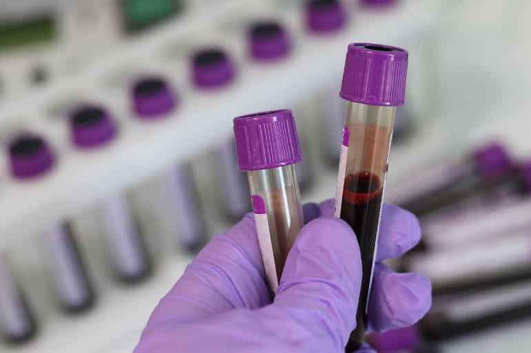
Together with neurologists at the Cantonal Hospital in St. Gallen, Nirmalraj has now successfully completed an initial study. For their pilot study, he examined blood samples from 50 patients and 16 healthy subjects. Using AFM technology, the Empa researcher analyzed the surface of around 1000 red blood cells per person without knowing anything about their state of health.
“This was the only way to make sure the interpretation of the data remained objective,” says Nirmalraj.
Protein fibers as an indicator
The Empa researcher measured the size, structure and texture of protein accumulations found on the blood cells. After thousands of red blood cells, the team eagerly awaited the comparison of the results from Nirmalraj’s counts with the clinical data from the neurologists. And indeed, the researchers were able to discern a pattern that matched the patients’ disease stage: People who had Alzheimer’s disease had large amounts of protein fibers made up of beta-amyloid peptides and tau proteins.

The proteins were able to assemble into fibers several hundred nanometers long. In healthy individuals or those with incipient brain disorders, however, Nirmalraj counted only few fibers.
This proves the feasibility of blood analysis using AFM technology, the Empa researcher says: “If a reliable blood test can be developed based on this method, people with suspected Alzheimer’s would be spared the unpleasant puncture of the spinal canal in order to be able to diagnose the disease reliably.”
However, there is still a long way to go before a simple blood test is available in hospitals. The team’s next step is to corroborate the data by studying a larger number of subjects at different stages of the disease using AFM and chemical analysis.
About this Alzheimer’s disease research news
Author: Peter Nirmalraj
Source: EMPA
Contact: Peter Nirmalraj – EMPA
Image: The image is in the public domain
Original Research: Open access.
“Spatial organization of protein aggregates on red blood cells as physical biomarkers of Alzheimer’s disease pathology” by Peter Nirmalraj et al. Science Advances
Abstract
Spatial organization of protein aggregates on red blood cells as physical biomarkers of Alzheimer’s disease pathology
Quantifying physical differences of protein aggregates implicated in Alzheimer’s disease (AD), in blood, could provide crucial information on disease stages. Here, red blood cells (RBCs) from 50 patients with neurocognitive complaints and 16 healthy individuals were profiled using an atomic force microscope (AFM).
AFM measurements revealed patient age– and stage of neurocognitive disorder–dependent differences in size, shape, morphology, assembly, and prevalence of protein aggregates on RBCs, referred to as physical biomarkers.
Crystals composed of fibrils were exclusively detected on RBCs for AD patients aged above 80 years. Fibril prevalence was negatively correlated with the cerebrospinal fluid (CSF) β-amyloid (Aβ) 42/40 ratio and was observed to be higher in the Aβ-positive patient category. Using a cutoff of ≥40% fibril prevalence, the CSF Aβ status was classified with 88% accuracy (sensitivity 100%, specificity 73%).
The merits and challenges in integrating physical biomarkers in AD diagnosis are discussed.


