

Summary: A new study sheds light on the neurobiology of reading.
Source: University of Pittsburgh.
Using direct neural recordings from the visual word form area, researchers were able to see words that patients read as the patients read them.
Reading is a relatively modern and uniquely human skill. For this reason, visual word recognition has been a puzzle for neuroscientists because the neural systems responsible for reading could not have evolved for this purpose. “The existence of brain regions dedicated to reading has been fiercely debated for almost 200 years,” said Avniel Ghuman, an assistant professor in the University of Pittsburgh Department of Neurological Surgery. “Wernicke, Dejerine, and Charcot, among the most important and influential neurologists and neuroscientists of the 19th century, debated whether or not there was a visual center for words in the brain.”
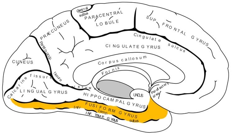
In recent years, much of this debate has centered on the left mid-fusiform gyrus, which some call the visual word form area. A recent study by Pitt neuroscience researchers addresses this debate and sheds light on our understanding of the neurobiology of reading.
In a study to be published July 19 in the Proceedings of the National Academy of Sciences, Ghuman, Elizabeth Hirshorn of Pitt’s Learning Research and Development Center (LRDC), and colleagues from the Department of Psychology and Center for the Neural Basis of Cognition used direct neural recordings and brain stimulation to study the role of the visual word form area in reading in four epileptic patients. The patients chose surgical treatment for their drug-resistant epilepsy and volunteered to participate in the research study. As part of the surgical treatment, neurosurgeons implanted electrodes in the patients’ visual word form area, providing an unprecedented opportunity to understand how the brain recognizes printed words.
First, painless electrical brain stimulation was used through the electrodes to disrupt the normal functioning of the visual word form area, which adversely affected the patients’ ability to read words. One patient dramatically misperceived letters, and another felt that there were words and parts of words present that were not in what she was reading. Stimulation to this region did not disrupt their ability to name objects or faces.
In addition to stimulating through these electrodes, the activity from the area was recorded while the patients read words. Using techniques from machine learning to analyze the brain activity that evolved over a few hundred milliseconds from this region, the researchers could tell what word a patient was reading at a particular moment. This suggests that neural activity in the area codes knowledge about learned visual words that can be used to discriminate even words that are only one letter different from one another (for example, “hint” and “lint”).
“This study shows that the visual word form area is exquisitely tuned to the fine details of written words and that this area plays a critical role in refining the brain’s representation of what we are reading. The disrupted word and letter perception seen with stimulation provides direct evidence that the visual word form area plays a dedicated role in skilled reading,” said Hirshorn. “These results also have important implications for understanding and treating reading disorders. The activity in the visual word form area, along with its interactions with other brain areas involved in language processing, could be a marker for proficient reading. Having a better understanding of this neural system could be critical for diagnosing reading disorders and developing targeted therapies.”
“It is exciting that with modern brain-recording techniques and advanced analysis methods, we are finally able to start answering questions about the brain and the mind that people have asked for centuries and contribute to our understanding of reading disorders,” said Ghuman.
Source: Joe Miksch – University of Pittsburgh
Image Source: This NeuroscienceNews.com image is in the public domain.
Video Source: The video is credited to Laboratory of Cognitive Neurodynamics.
Original Research: Full open access research for “Decoding and disrupting left midfusiform gyrus activity during word reading” by Elizabeth A. Hirshorn, Yuanning Li, Michael J. Ward, R. Mark Richardson, Julie A. Fiez, and Avniel Singh Ghuman in PNAS. Published online July 19 2016 doi:10.1073/pnas.1604126113
[cbtabs][cbtab title=”MLA”]University of Pittsburgh. “How Words Are Represented in the Brain.” NeuroscienceNews. NeuroscienceNews, 21 July 2016.
<https://neurosciencenews.com/visual-word-form-area-neuroscience-4723/>.[/cbtab][cbtab title=”APA”]University of Pittsburgh. (2016, July 21). How Words Are Represented in the Brain. NeuroscienceNew. Retrieved July 21, 2016 from https://neurosciencenews.com/visual-word-form-area-neuroscience-4723/[/cbtab][cbtab title=”Chicago”]University of Pittsburgh. “How Words Are Represented in the Brain.” https://neurosciencenews.com/visual-word-form-area-neuroscience-4723/ (accessed July 21, 2016).[/cbtab][/cbtabs]
Abstract
Decoding and disrupting left midfusiform gyrus activity during word reading
The nature of the visual representation for words has been fiercely debated for over 150 y. We used direct brain stimulation, pre- and postsurgical behavioral measures, and intracranial electroencephalography to provide support for, and elaborate upon, the visual word form hypothesis. This hypothesis states that activity in the left midfusiform gyrus (lmFG) reflects visually organized information about words and word parts. In patients with electrodes placed directly in their lmFG, we found that disrupting lmFG activity through stimulation, and later surgical resection in one of the patients, led to impaired perception of whole words and letters. Furthermore, using machine-learning methods to analyze the electrophysiological data from these electrodes, we found that information contained in early lmFG activity was consistent with an orthographic similarity space. Finally, the lmFG contributed to at least two distinguishable stages of word processing, an early stage that reflects gist-level visual representation sensitive to orthographic statistics, and a later stage that reflects more precise representation sufficient for the individuation of orthographic word forms. These results provide strong support for the visual word form hypothesis and demonstrate that across time the lmFG is involved in multiple stages of orthographic representation.
“Decoding and disrupting left midfusiform gyrus activity during word reading” by Elizabeth A. Hirshorn, Yuanning Li, Michael J. Ward, R. Mark Richardson, Julie A. Fiez, and Avniel Singh Ghuman in PNAS. Published online July 19 2016 doi:10.1073/pnas.1604126113


