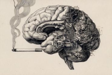
Adolescence is a dangerous time for the onset of mental health problems. Advances in brain imaging are helping to picture how neural changes in these crucial years can lead to chronic debilitating mental illness.
Restless, disordered, uncertain, impulsive, emotional – the teenage brain can be a confused fury of neural firings and misfirings.
For most 14- to 24-year-olds – the “risky age” as Professor Ed Bullmore describes it – the maelstrom eventually subsides. For some, episodes of depression, low self-esteem, self-harm or paranoia may intensify and become more frequent. For around 1 in 100, the change in mental state is so marked that it will become difficult for them to distinguish their delusions and hallucinations from reality – one of the hallmarks of schizophrenia.
“Schizophrenia is a particularly feared diagnosis,” says Bullmore. “People tend to think it means a chronic lifelong dependency on medication and therapy. It can mean this, but it can also last only a few years. The main thing that patients and their families want to know is what does the future hold – am I likely to be able to resume my life, get a job, and so on?”
Bullmore is co-chair of Cambridge Neuroscience, an initiative to enhance multidisciplinary research across the University, and leads the Department of Psychiatry, where he and colleagues have been developing imaging techniques that are revealing where and over what timescale abnormalities in the brain develop in people with mental health problems.
This is no easy task. Even being able to show a neural abnormality has been a major and relatively recent advance for understanding a condition that, Bullmore says, has in the past been regarded with prejudice and assumptions. “Demonstrating neural change moves us away from what might be regarded as a blaming approach where someone is made to feel personally responsible for the fact these symptoms exist. Imaging shows you that’s not the case – there is a biological basis.”
The task is made difficult because there is no single event or area of the brain that underlies schizophrenia. It has only been from the collation of results from imaging studies worldwide that it has become apparent that when it comes to mental health disorders the scientists need to look at the big picture – the changes happening in wiring circuits across the whole brain.
Imaging techniques such as magnetic resonance imaging (MRI) are helping to map the brain in unprecedented detail. Structural MRI follows the movement of water as it diffuses along the pathways forged by neurons – showing the network of connections spread across the brain. Functional MRI measures slow rhythmic activity in the brain; if two areas of the brain show activity at the same time the chances are they are functionally connected. Bullmore and colleagues have developed mathematical methods to calculate the probability of there being such a connection.
“Neuroscience is no longer just about neurons,” he explains. “We can also now talk in terms of hubs, networks and connectomes. If the brain is thought of as a computer, with ‘processors’ in the outer grey matter and ‘wires’ that connect them in the inner white matter, some hub regions are more highly connected than others.”
In schizophrenia, connectivity in the wiring diagram goes awry and highly connected hubs are especially affected – “you could call it a hubopathy,” says Bullmore. His team’s research has demonstrated that those who have suffered decades of schizophrenia have large-scale network abnormalities compared with a healthy brain, which goes some way to explaining the diversity and severity of symptoms experienced in schizophrenia. The question is: can imaging be used to chart this progression?
Bullmore and his colleagues believe so: “Roughly a third of patients recover, a third have intermittent symptoms and a third will be affected for decades by schizophrenia. At diagnosis we can’t currently tell which of these outcomes lies in store. But we think one day we will be able to correlate the pattern of network activity with future outcome.”
It’s not only what happens to patients post-diagnosis that interests Bullmore, but also what has happened neurologically in the years before diagnosis.
“For me, one of the most exciting aspects of psychiatry is that we can use imaging to study the ‘risky age’ of brain development to understand how the connectome grows or matures in healthy brains. We can then start to pinpoint which genetic and environmental factors might favour healthy adolescent brain network development and which factors might predispose to abnormal network development, leading to chronic disability or distress.”
In 2012, Bullmore and colleagues Professor Ian Goodyer and Professor Peter Jones in Cambridge’s Department of Psychiatry (in collaboration with Professor Ray Dolan and Professor Peter Fonagy from University College London) launched the NeuroScience in Psychiatry Network, funded by the Wellcome Trust. They have been recruiting a panel of 2,000 healthy volunteers aged 14–24 years, 300 of whom have had brain scans to contribute to one of the most comprehensive ‘circuit diagrams’ of the teenage brain ever attempted.
“Remarkably little is known about how brain networks grow during the crucial transition from childhood dependence to life as independent adults,” adds Bullmore. “The adolescent brain is still a bit of a black box. But it is a big step forward that we can now see healthy human brain development much more clearly, especially with the next-generation brain scanners coming to Cambridge soon [see panel]. It’s very exciting to think that we should then be able to understand and predict the pathways of brain network development that lead to schizophrenia.”
Opening the black box
The arrival of two state-of-the-art MRI machines in Cambridge, thanks to funding from the Medical Research Council (MRC), will revolutionise the study of the brain.

“A brain scan is much more than an image,” contends Ed Bullmore. “It’s really a very large collection of numbers. With the best scanners and some high-performance computing, you can start to think not only about disease mechanisms but also about identifying early risk factors and preventative action.”
Two of the newest scanners in the UK will arrive at the Cambridge Biomedical Campus in 2016, and a new high-speed secure link will be created through to the recently opened £20 million West Cambridge Data Centre, which will analyse the data.
One scanner, a new 7-Tesla ‘ultrahigh-field’ MRI machine, will help researchers see how the human brain works as a whole, yet also with the precision of a grain of sand a fraction of a millimetre across. It will further the study of dementia, brain injury, obesity, addiction, mental health disorders, pain and stroke.
‘7T’ is a collaboration between the University and the MRC’s Cognition and Brain Sciences Unit (CBSU). Professor James Rowe, from the Department of Clinical Neurosciences and the CBSU, explains: “The new scanner is a major advance to study the details of the human brain not only in health but also the effects of age and the origins of brain diseases. The unprecedented detail and sensitivity at 7T is essential in the national effort towards a cure for dementia and mental illness.”
Joining the 7T scanner will be a positron emission tomography (PET)–MRI machine, which shows changes in the brain down to the level of individual molecules. Until now only two PET–MRI scanners existed in the UK, but MRC Dementias Platform UK has invested in five more nationally, creating what is thought to be the first nationally coordinated MRI–PET network anywhere in the world.
“Beating dementia is a long-term goal,” adds Rowe. “These scanners will make a very significant contribution to this eventual success and to the lives of patients and their families.”
Source: Louise Walsh – University of Cambridge
Image Credit: The image is credited to The District and is in the public domain.


